Japanese
English
症例
Ossified choroid plexus papillomaの1例
Ossified Choroid Plexus Papilloma: Case report
川俣 貴一
1
,
久保 長生
1
,
河村 弘庸
1
,
岩田 幸也
1
,
加川 瑞夫
1
,
喜多村 孝一
1
Takakazu KAWAMATA
1
,
Osami KUBO
1
,
Hirotsune KAWAMURA
1
,
Yukiya IWATA
1
,
Mizuo KAGAWA
1
,
Koichi KITAMURA
1
1東京女子医科大学脳神経センター脳神経外科
1Department of Neurosurgery, Neurological Institute, Tokyo Women's Medical College
キーワード:
Choroid Nexus hahilloma
,
Ossification
,
Calcification
,
Brain stone
Keyword:
Choroid Nexus hahilloma
,
Ossification
,
Calcification
,
Brain stone
pp.989-994
発行日 1988年7月10日
Published Date 1988/7/10
DOI https://doi.org/10.11477/mf.1436202671
- 有料閲覧
- Abstract 文献概要
- 1ページ目 Look Inside
I.はじめに
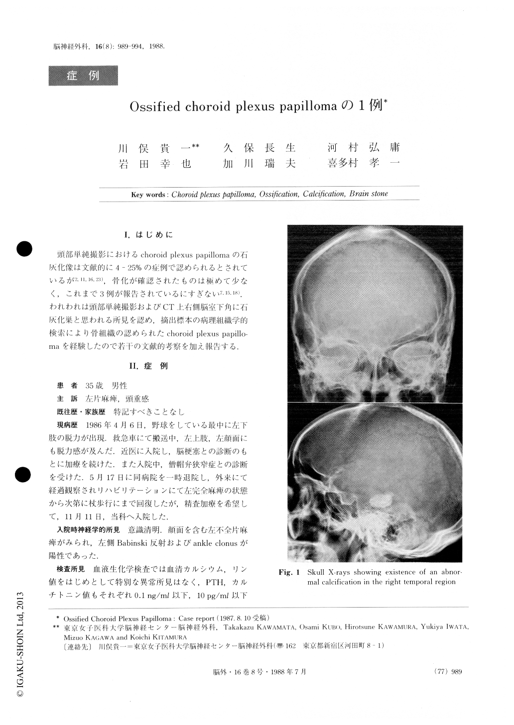
頭部単純撮影におけるchoroid plexus papillomaの石灰化像は文献的に4-25%の症例で認められるとされているが2,11,16,23),骨化が確認されたものは極めて少なく,これまで3例が報告されているにすぎない7,15,18).われわれは頭部単純撮影およびCT—上右側脳室下角に石灰化巣と思われる所見を認め,摘出標本の病理組織学的検索により骨組織の認められたchcoroid plexus papillo—maを経験したので若干の文献的考察を加え報告する.
While the calcification has been documented radiolo-gically in 4 - 25 percent of the cases with choroid plex-us papilloma, the ossification of choroid plexus papillo-ma has been reported only in 3 cases on literature. In this paper, we present a case of large ossified choroid plexus papilloma in the right lateral ventricle.
A 35-year-olcl man was admitted with left hemi-paresis and headache. Skull X-rays showed an abnor-mal calcified mass (25 mm × 23 mm × 14 mm) in the right temporal region.
Copyright © 1988, Igaku-Shoin Ltd. All rights reserved.