Japanese
English
- 有料閲覧
- Abstract 文献概要
- 1ページ目 Look Inside
I.はじめに
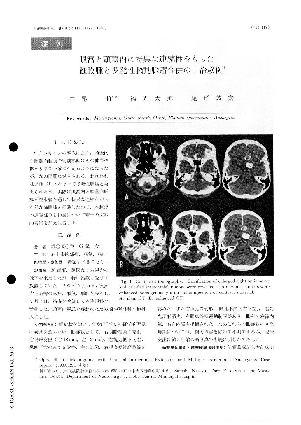
CTスキャンの導入により,頭蓋内や眼窩内腫瘍の術前診断はその伸展や拡がりまで正確に行えるようになったが,なお困難な場合もある,われわれは術前CTスキャンで多発性腫瘍と考えられたが,実際は眼窩内と頭蓋内腫瘍が視束管を通して特異な連続を持った稀な髄膜腫を経験したので,本腫瘍の原発部位と伸展について若干の文献的考察を加え報告する.
A 67-year-old houswife was admitted to our clinic who had a 37-year history of right visual impairment. On examination, conjunctival hyperemia, decreased visual acuity (light perception), proptosis (6mm), optic artophy, irregularity of the pupil and slight impairment of abduction were revealed in her right eve. She had no other neurological dificits. Routine x-ray of the skull and orbits showed shell-like calcifications at anterior cranial fossa and serpentine calcified mass in the right orbit. Tomograms of the orbits indicated slight enlargement of the right optic canal, but did not showed any abnormalities in the orbital walls and roofs.

Copyright © 1981, Igaku-Shoin Ltd. All rights reserved.


