Japanese
English
- 有料閲覧
- Abstract 文献概要
- 1ページ目 Look Inside
- 参考文献 Reference
Ⅰ.はじめに
Reversible posterior leukoencephalopathy syndrome (RPLS)は,1996年,Hincheyら3)が後頭葉優位の白質病変を主とし,頭痛,痙攣,意識障害といった共通の臨床症候と画像所見を呈する疾患群を1つの概念としてまとめて提唱したものである.今回,われわれはこの画像所見にほぼ合致する3例を経験した.しかし,臨床症状やその転帰はさまざまであり,この概念の問題点も含めて報告する.
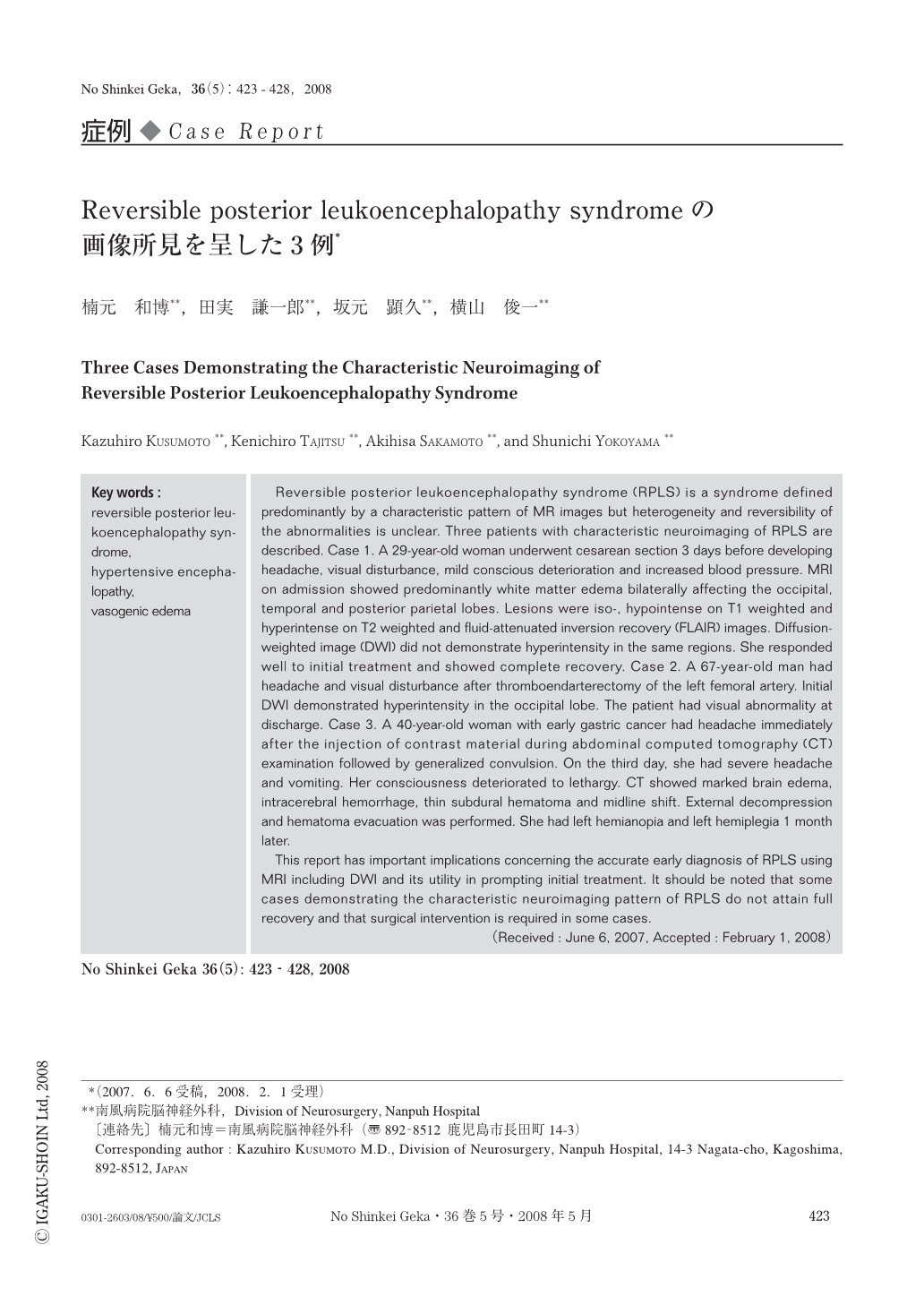
Reversible posterior leukoencephalopathy syndrome (RPLS) is a syndrome defined predominantly by a characteristic pattern of MR images but heterogeneity and reversibility of the abnormalities is unclear. Three patients with characteristic neuroimaging of RPLS are described. Case 1. A 29-year-old woman underwent cesarean section 3 days before developing headache, visual disturbance, mild conscious deterioration and increased blood pressure. MRI on admission showed predominantly white matter edema bilaterally affecting the occipital, temporal and posterior parietal lobes. Lesions were iso-, hypointense on T1 weighted and hyperintense on T2 weighted and fluid-attenuated inversion recovery (FLAIR) images. Diffusion-weighted image (DWI) did not demonstrate hyperintensity in the same regions. She responded well to initial treatment and showed complete recovery. Case 2. A 67-year-old man had headache and visual disturbance after thromboendarterectomy of the left femoral artery. Initial DWI demonstrated hyperintensity in the occipital lobe. The patient had visual abnormality at discharge. Case 3. A 40-year-old woman with early gastric cancer had headache immediately after the injection of contrast material during abdominal computed tomography (CT) examination followed by generalized convulsion. On the third day, she had severe headache and vomiting. Her consciousness deteriorated to lethargy. CT showed marked brain edema, intracerebral hemorrhage, thin subdural hematoma and midline shift. External decompression and hematoma evacuation was performed. She had left hemianopia and left hemiplegia 1 month later.
This report has important implications concerning the accurate early diagnosis of RPLS using MRI including DWI and its utility in prompting initial treatment. It should be noted that some cases demonstrating the characteristic neuroimaging pattern of RPLS do not attain full recovery and that surgical intervention is required in some cases.

Copyright © 2008, Igaku-Shoin Ltd. All rights reserved.


