Japanese
English
- 有料閲覧
- Abstract 文献概要
- 1ページ目 Look Inside
Ⅰ.はじめに
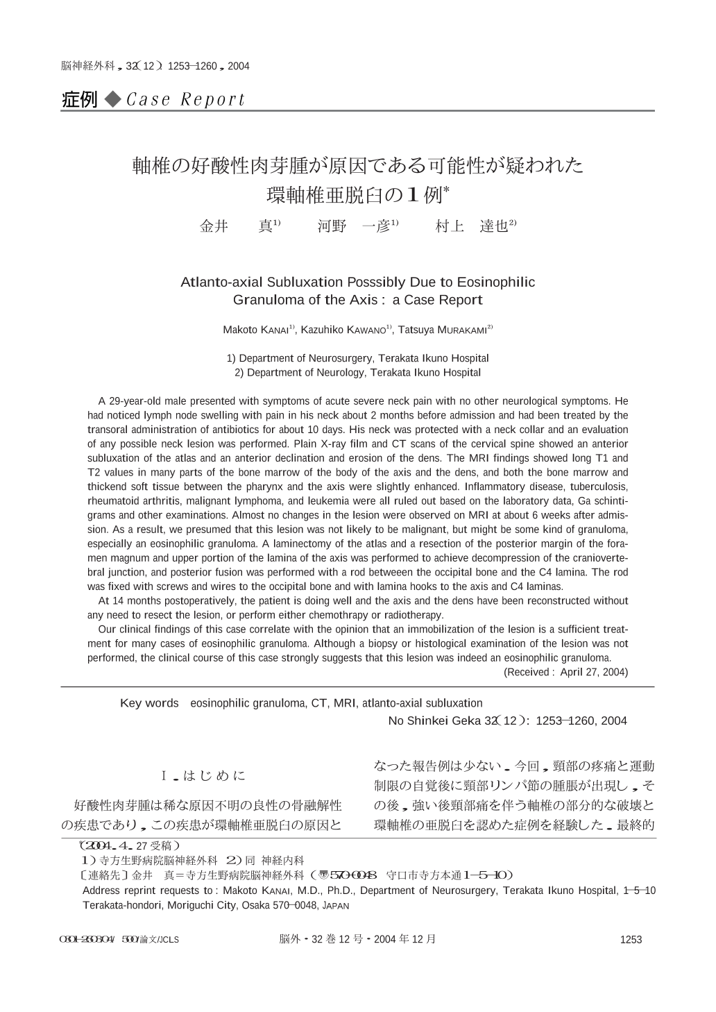
好酸性肉芽腫は稀な原因不明の良性の骨融解性の疾患であり,この疾患が環軸椎亜脱臼の原因となった報告例は少ない.今回,頸部の疼痛と運動制限の自覚後に頸部リンパ節の腫脹が出現し,その後,強い後頸部痛を伴う軸椎の部分的な破壊と環軸椎の亜脱臼を認めた症例を経験した.最終的に好酸性肉芽腫が原因である可能性も考え,第1頸髄レベルでの頸髄の後方からの除圧と後頭骨から第4頸椎レベルまでの後方固定術を施行し,良好な結果を得たので報告する.
A 29-year-old male presented with symptoms of acute severe neck pain with no other neurological symptoms. He had noticed lymph node swelling with pain in his neck about 2 months before admission and had been treated by the transoral administration of antibiotics for about 10 days. His neck was protected with a neck collar and an evaluation of any possible neck lesion was performed. Plain X-ray film and CT scans of the cervical spine showed an anterior subluxation of the atlas and an anterior declination and erosion of the dens. The MRI findings showed long T1 and T2 values in many parts of the bone marrow of the body of the axis and the dens,and both the bone marrow and thickend soft tissue between the pharynx and the axis were slightly enhanced. Inflammatory disease,tuberculosis,rheumatoid arthritis,malignant lymphoma,and leukemia were all ruled out based on the laboratory data,Ga schintigrams and other examinations. Almost no changes in the lesion were observed on MRI at about 6 weeks after admission. As a result,we presumed that this lesion was not likely to be malignant,but might be some kind of granuloma,especially an eosinophilic granuloma. A laminectomy of the atlas and a resection of the posterior margin of the foramen magnum and upper portion of the lamina of the axis was performed to achieve decompression of the craniovertebral junction,and posterior fusion was performed with a rod betweeen the occipital bone and the C4 lamina. The rod was fixed with screws and wires to the occipital bone and with lamina hooks to the axis and C4 laminas.
At 14 months postoperatively,the patient is doing well and the axis and the dens have been reconstructed without any need to resect the lesion,or perform either chemothrapy or radiotherapy.
Our clinical findings of this case correlate with the opinion that an immobilization of the lesion is a sufficient treatment for many cases of eosinophilic granuloma. Although a biopsy or histological examination of the lesion was not performed,the clinical course of this case strongly suggests that this lesion was indeed an eosinophilic granuloma.

Copyright © 2004, Igaku-Shoin Ltd. All rights reserved.


