Japanese
English
- 有料閲覧
- Abstract 文献概要
- 1ページ目 Look Inside
- 参考文献 Reference
Ⅰ.はじめに
脳神経外科領域では,手術をより確実で安全なものとするために少しでも多くの情報を得ようと,形態のみならず機能をも可視化する画像診断法が積極的に導入されてきた.最近は,術中ナビゲーションシステムに解剖や機能に関する様々な画像情報を統合したfunctional neuronavigationも汎用されてきており,脳神経外科手術自体が画像情報を駆使したスタイルへと変遷を遂げている.
新規画像診断技術の開発に関して脳神経外科医の立場から言えば,顕微鏡を駆使した繊細な手術を手掛けるがために「さらに詳細な解剖を知りたい」,またquality of lifeにかかわる脳機能の温存に努めるがために「直接見ることのできない機能を可視化したい」という2つの大きな欲求が根底にある.前者に対しては,3.0T装置をはじめとするMRIの高磁場化や3次元画像構築技術の進歩があり,後者に対しては functional MRI(fMRI),positron emission tomography(PET),magnetoencephalography(MEG)をはじめとした機能画像法の開発や,diffusion tensor imaging(DTI)による白質情報の画像化,MR spectroscopy(MRS)のような代謝性産物の分子イメージングなどの発展があった.これら新規画像診断法を臨床の中で運用していくにあたって,脳神経外科医は唯一脳そのものに触れることができる立場にあるものとして,術中モニタリングや頭蓋内マッピングなどの神経生理学的手法での検証や,摘出標本の病理学的所見との比較に絶えず取り組み,また新たな画像法開発のためにその結果をフィードバックしていかなくてはならない.
われわれは,脳研究所所属の脳神経外科学教室として,他の脳研究部門との連携が自由にある環境を生かし注1),今まで「脳腫瘍」「脳血管障害」「機能性疾患」などの各脳神経外科領域に適応すべき高解像度解剖画像や機能画像診断法の確立,つまり脳神経外科領域での画像診断法において一貫したtranslational researchに取り組んできた.本報告では,MRI画像の研究に関しては新潟大学統合脳機能研究センターと,MEGとてんかんの研究に関しては国立病院機構西新潟中央病院てんかんセンターと,optical imagingに関しては新潟大学脳研究所システム脳生理学教室とともに手掛けて来た研究を中心に,脳神経外科領域における新規画像診断について現在のトピックと今後の展望を交えて紹介する.
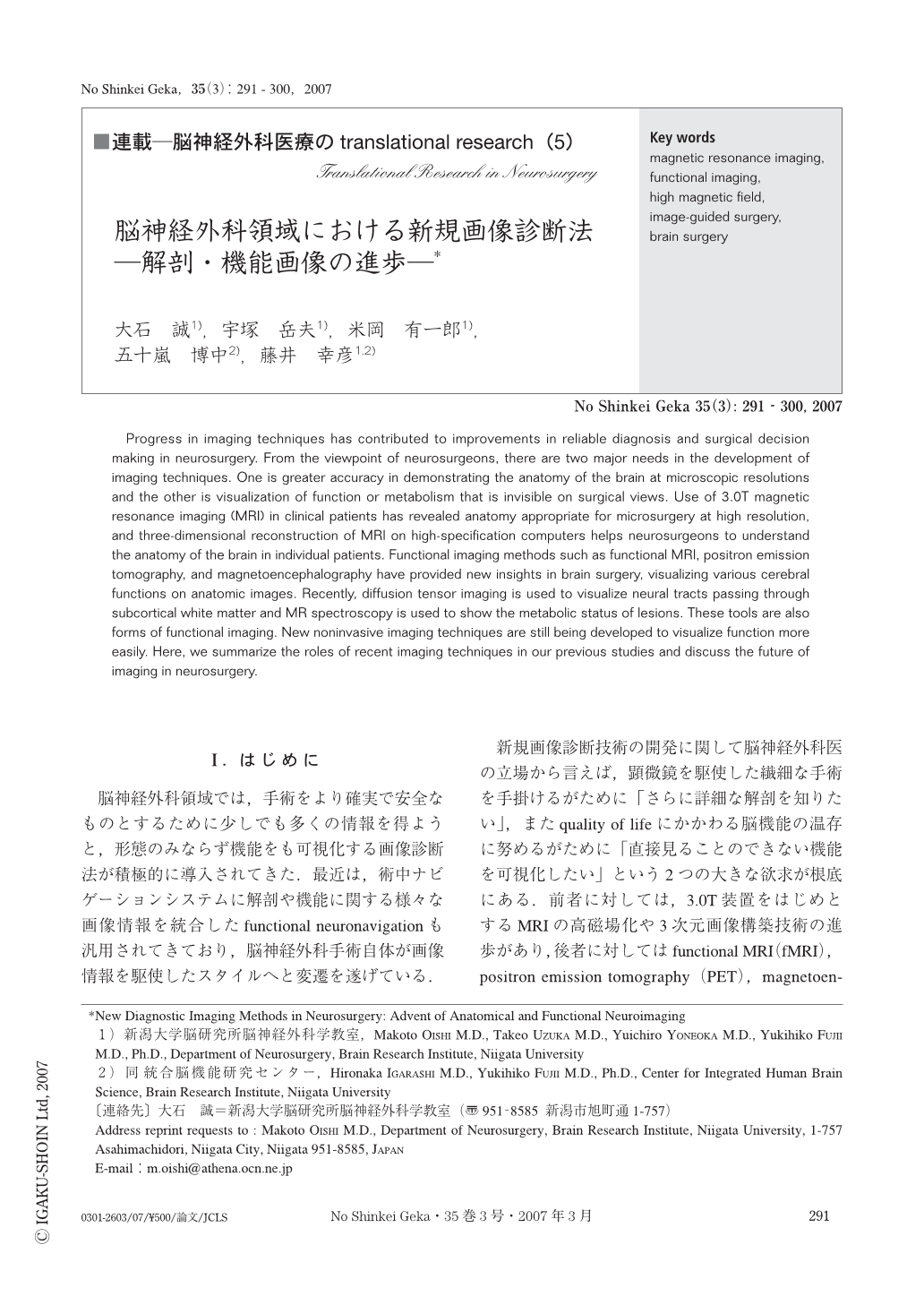
Progress in imaging techniques has contributed to improvements in reliable diagnosis and surgical decision making in neurosurgery. From the viewpoint of neurosurgeons, there are two major needs in the development of imaging techniques. One is greater accuracy in demonstrating the anatomy of the brain at microscopic resolutions and the other is visualization of function or metabolism that is invisible on surgical views. Use of 3.0T magnetic resonance imaging (MRI) in clinical patients has revealed anatomy appropriate for microsurgery at high resolution, and three-dimensional reconstruction of MRI on high-specification computers helps neurosurgeons to understand the anatomy of the brain in individual patients. Functional imaging methods such as functional MRI, positron emission tomography, and magnetoencephalography have provided new insights in brain surgery, visualizing various cerebral functions on anatomic images. Recently, diffusion tensor imaging is used to visualize neural tracts passing through subcortical white matter and MR spectroscopy is used to show the metabolic status of lesions. These tools are also forms of functional imaging. New noninvasive imaging techniques are still being developed to visualize function more easily. Here, we summarize the roles of recent imaging techniques in our previous studies and discuss the future of imaging in neurosurgery.

Copyright © 2007, Igaku-Shoin Ltd. All rights reserved.


