Japanese
English
- 有料閲覧
- Abstract 文献概要
- 1ページ目 Look Inside
I.はじめに
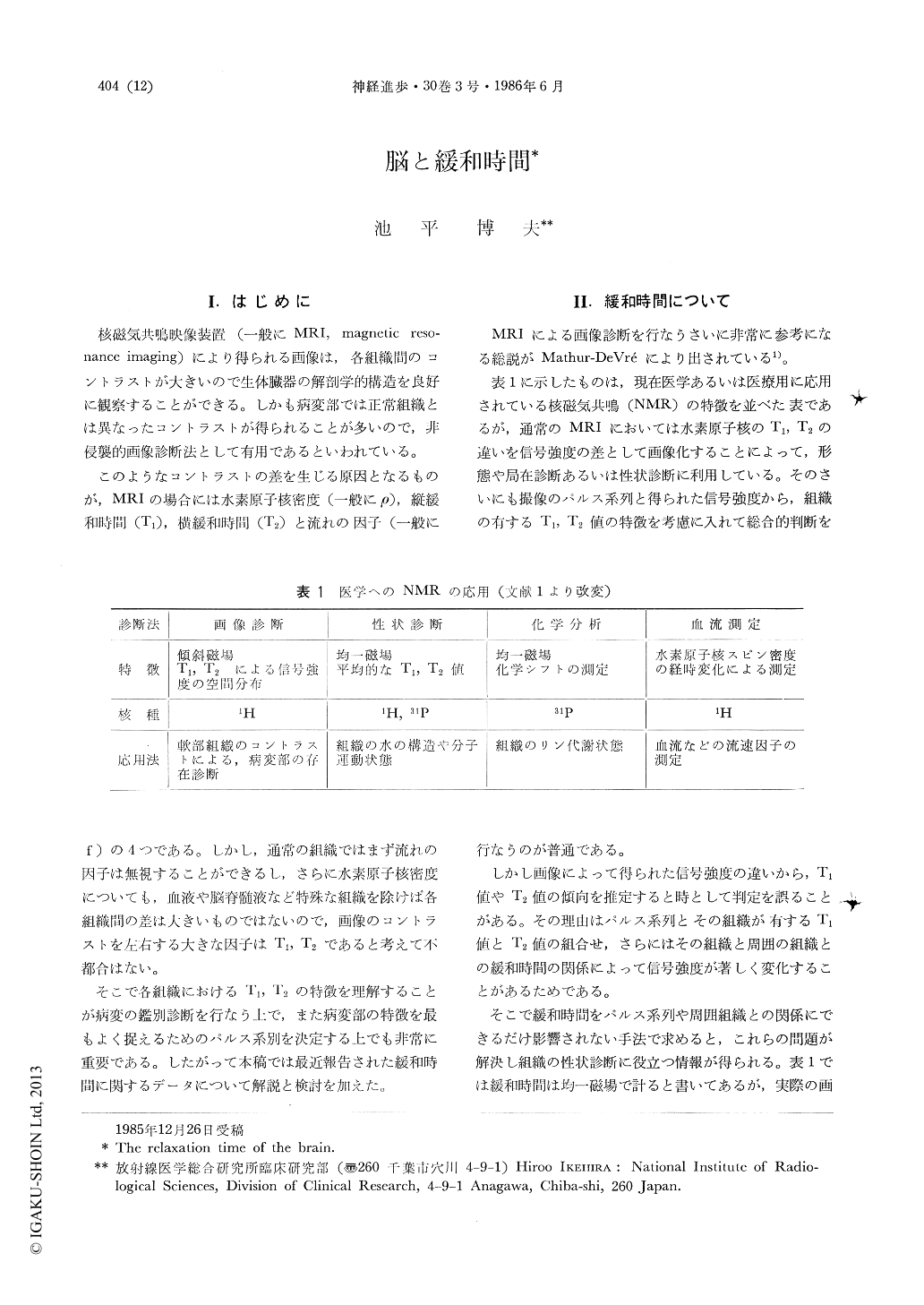
核磁気共鳴映像装置(一般にMRI,magnetic resonance imaging)により得られる画像は,各組織間のコントラストが大きいので生体臓器の解剖学的構造を良好に観察することができる。しかも病変部では正常組織とは異なったコントラストが得られることが多いので,非侵襲的画像診断法として有用であるといわれている。
このようなコントラストの差を生じる原因となるものが,MRIの場合には水素原子核密度(一般にρ),縦緩和時間(T1),横緩和時間(T2)と流れの因子(一般にf)の4つである。しかし,通常の組織ではまず流れの因子は無視することができるし,さらに水素原子核密度についても,血液や脳脊髄液など特殊な組織を除けば各組織間の差は大きいものではないので,画像のコントラストを左右する大きな因子はT1,T2であると考えて不都合はない。
Over past a couple of years, many reports about the relation between T1 (longitudinal relaxation time) or T2 (transverse relaxation time) and the human normal and abnormal tissues, were published by many researchers of all over the world.
These T1 or T2 data are almost decided for normal or some basic pathological tissues, so if we know these data, it will be very useful for the differential diagnosis using MRI machine, and we are able to understand the limit of the differential diagnosis using MRI, also we are possible to use MRI machine most effectively.

Copyright © 1986, Igaku-Shoin Ltd. All rights reserved.


