Japanese
English
- 有料閲覧
- Abstract 文献概要
- 1ページ目 Look Inside
I.まえがき
脳の血管性障害においてもつとも問題になることの一つはその後遺症であり,後遺症としては片麻痺がもつとも問題であるが,しばしばrehabilitationと関係して言語障害,ことに失語症も問題となる。近年われわれの研究でも明らかにされたように,わが国においては脳出血に比べて脳血栓が明らかに増加しつつあるという事実がある。この脳血栓においては発作時の危機を脱して生残る人が多く,したがつて後遺症は今日われわれがもつとも関心を有する問題である。
失語症は従来優位半球の障害に基づくとされているが,その優位半球とは利き手を支配する半球であると考えられており,これは今日の常識である。たとえば大橋29)の各種の脳障害200例についての半球症状をみると,左半球の障害では失語症がもつとも多数をしめ,その他に失読,失書,失算などの言語と関係する症状,その他失行,失認が記載されている。右半球の障害においては,着衣失行,半側空間および身体失認,地誌的失見当が多く現われている。またLuria23)らの特定の機能についてこれを支配する脳半球をもつて優位半球とする考えもある。
The purpose of this paper is to show the studiesof some functional impairments in cerebrovasculardisorders, particularly in reference to the dominanthemisphere.
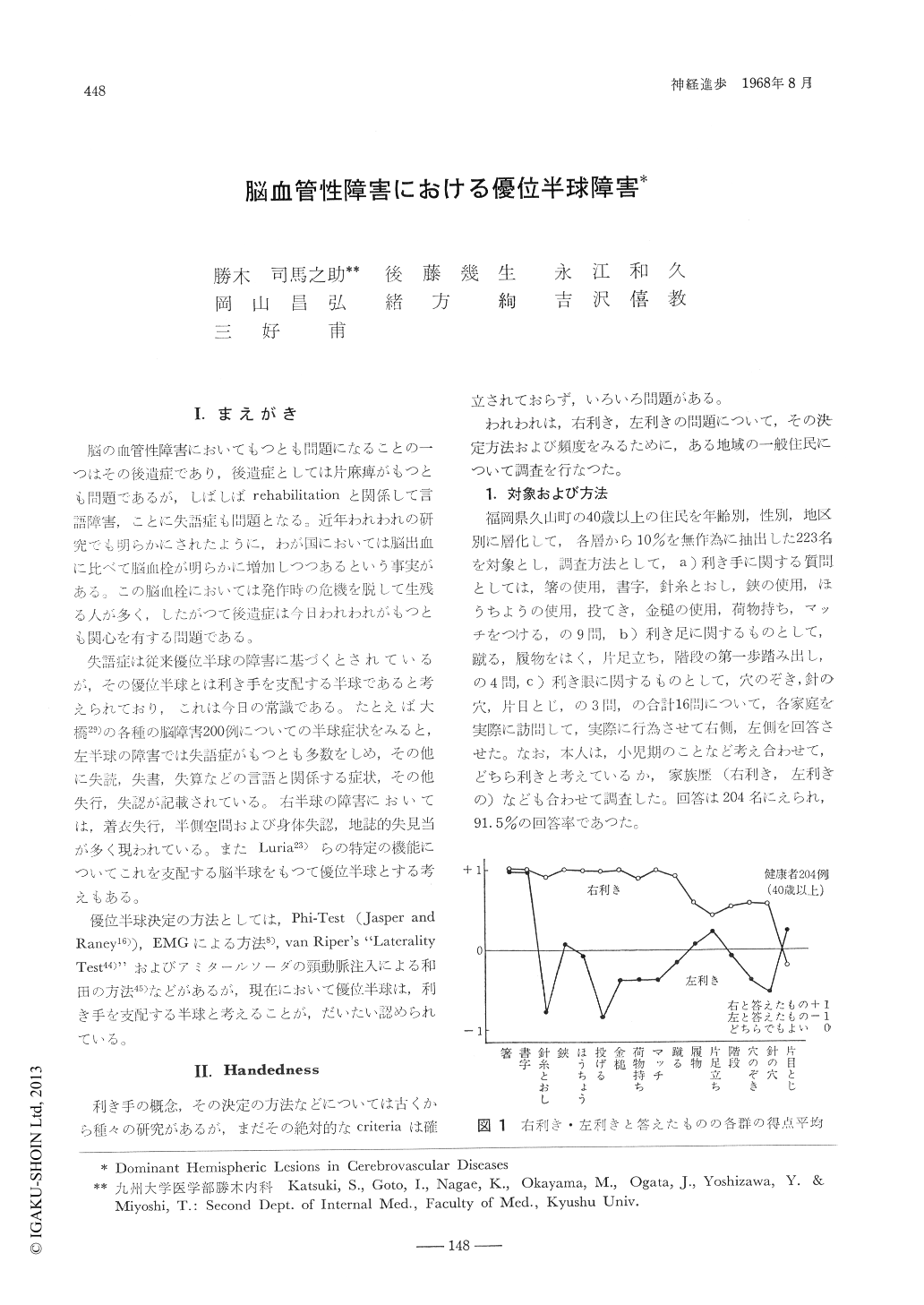
Two hundred and four residents of Hisayamaselected by stratified random sampling's method,all over 40 years old, were investigated by 16different questions or tasks to determine the pre-ference for hand, foot and eye.
Thirteen left-hand edsubjects (6.4%) were foundin this study. The activities such as "throw anobject" and "thread a needle" had a good corre-lation to the manual superiority, while the activi-ties such as "write" and "eat with chopsticks" inleft-handed people were found to have convertedto the right side in all cases. There are no closerelationships among handedness, foot superiorityand eye-superiority.
It is known that some abnormalities in psycho-metric tests and in tests for apraxia, agnosia andaphasia are very often recognized in cerebrallesions. Therefore, tests for apraxia, agnosia andaphasia and several psychometric tests were givenin cerebrovascular disease (CVD) cases: tests forapraxia, agnosia and aphasia in 197, WechslerAdult Intelligence Scales (WAIS) in 63, PorteusMaze Test in 46, Trail Making Test in 49 andBender Gestalt Test in 110 cases. The patientswith right hemispheric lesions showed poor resultsin the Perfbrmance Test of WAIS, the Trail Ma-king Test and the Bender Gestalt Test, whilethe patients with left hemispheric lesions showedpoor results in both Verbal and Perliirmance Testsof WAIS. Even poorer results in these tests werefound in the patients with severe cerebral lesionsin comparison to mild cerebral damages.
Apraxia and agnosia were noted in only 3 of197 GVD cases (1. 5%), while aphasia was foundin 47 of 197 CVD cases (24%) and in 45 of 81right hemiplegics (56%), in 13 of 13 right hemi-plegic cases with cerebral hemorrhage (100%) andin 24 of 58 right herniplegic cases with cerebral thrombosis (41%).
There was a close relationship between theclinical severity and the severity of aphasia, evalu-ated by the .Iapanesc version of Schuell's tests.
Sixty four of 73 CVD cases with EEG abnor-mality in the left cerebral hemisphere were aphasic. Aphasia, however, was found in only 2of 60 cases with EEG abnormalities in the rightcerebral hemisphere; one of which was a left-handed case and the other was shown by varioustests to be a crossed aphasic subject. Localizedabnormalities in EEG were found primarily in thetemporal, frontal and/or central lobe areas ofthe dominant hemisphere.
On angiographic examination, stenotic findingsof the internal carotid artery in the dominanthemispheric side were demonstrated in 10 of 14aphasics and in 4 of 16 non-aphasic patients.Severe aphasia was found in all 5 cases with com-plete occulsion of the left middle cerebral arterybefore ramification of the striate arteries, whilefive cases with complete occulsion of the leftmiddle cerebral artery after ramification of thestriate arteries, however, did not show such asevere aphasic manifestation.
Pathoanatomical studies were also performed in11 cases of cerebral infarction with aphasia, one ofwhich was left-handed. Main cerebral lesions inall 11 cases were located in the areas suppliedby the middle cerebral artery in the dominanthemisphere.
In 2 mild aphasic cases one had lesions of thelateral portion of the right basal ganglia and theinsular cortex and the other had lesions sf thecortex and subjacent white matter of the leftmiddle and inferior frontal gyri.
In 9 severe aphasic cases, cerebral lesions occur-red as follows: in 2 cases they were located inthe area supplied by the left middle cerebralartery inclusive of the left basal ganglia, in 1case in the area supplied by the left middlecerebral artery exclusive of the basal ganglia, in 1case in the white matter of the area supplied bythe left middle cerebral artery, in 3 cases in thearea supplied by the left anterior and middlecerebral arteries, and in 2 cases in the parietaland temporal lobes inclusive of the basal gangliaand insular cortex. All 3 eases with occulsionthe origin of the left middle cerebral artery showedsevere aphasia.
Our results suggest that various psychometrictests and tests fUr apraxia, agnosia and aphasiaas well as EEG and cerebral angiography areuseful in determining the laterality and the extentof cerebral hemispheric lesions and also the severityof the brain damage.

Copyright © 1968, Igaku-Shoin Ltd. All rights reserved.


