Japanese
English
- 有料閲覧
- Abstract 文献概要
- 1ページ目 Look Inside
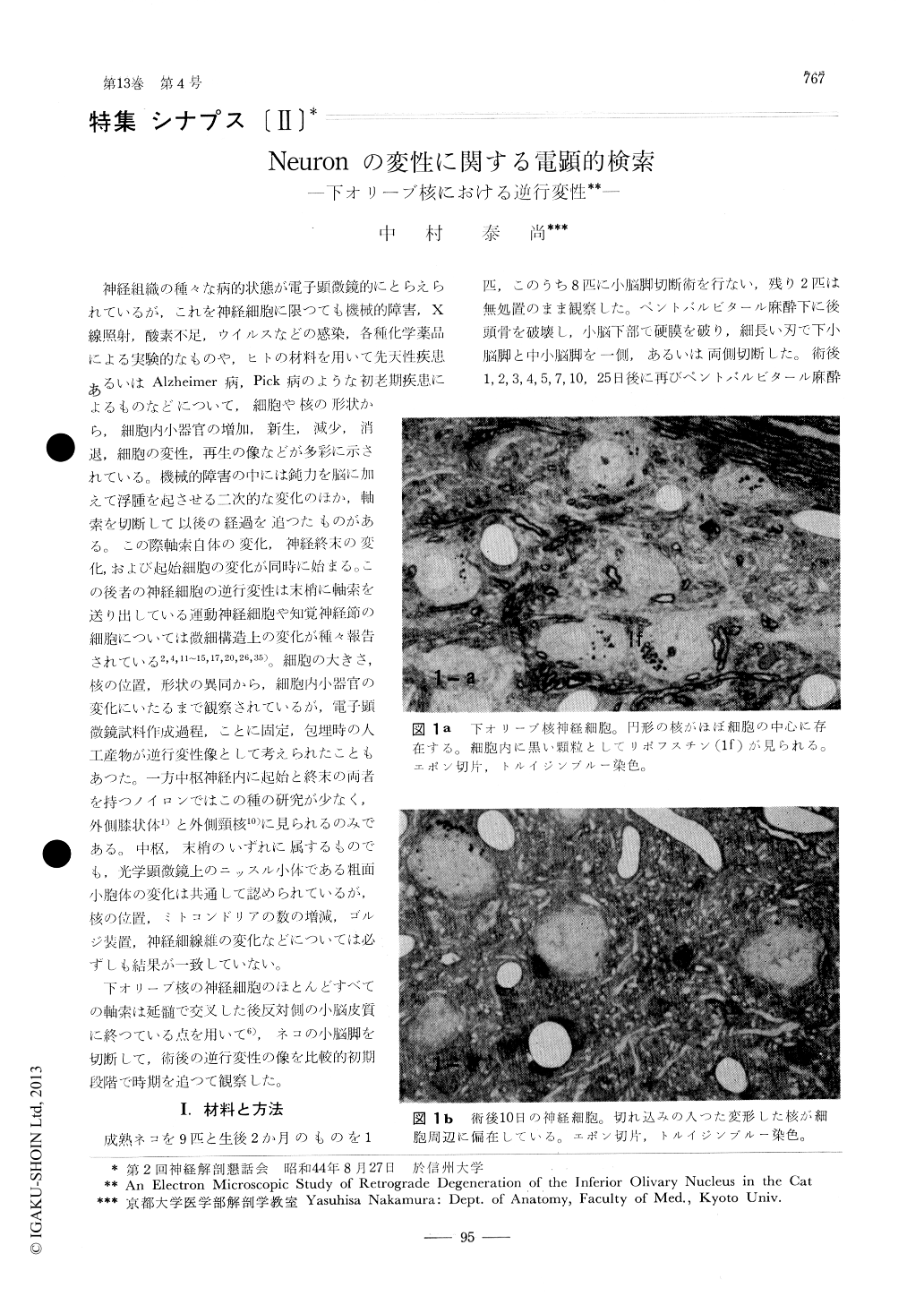
神経組織の種々な病的状態が電子顕微鏡的にとらえられているが,これを神経細胞に限つても機械的障害,X線照射,酸素不足,ウイルスなどの感染,各種化学薬品による実験的なものや,ヒトの材料を用いて先天性疾患あるいはAlzheimer病,Pick病のような初老期疾患によるものなどについて,細胞や核の形状から,細胞内小器官の増加,新生,減少,消退,細胞の変性,再生の像などが多彩に示されている。機械的障害の中には鈍力を脳に加えて浮腫を起させる二次的な変化のほか,軸索を切断して以後の経過を追つたものがある。この際軸索自体の変化,神経終末の変化,および起始細胞の変化が同時に始まる。この後者の神経細胞の逆行変性は末梢に軸索を送り出している運動神経細胞や知覚神経節の細胞については微細構造上の変化が種々報告されている2,4,11〜15,17,20,26,35)。細胞の大きさ,核の位置,形状の異同から,細胞内小器官の変化にいたるまで観察されているが,電子顕微鏡試料作成過程,ことに固定,包埋時の人工産物が逆行変性像として考えられたこともあつた。
Retrograde degeneration of the inferior olivary nucleus was produced by transection of the middle and inferior cerebellar peduncles. The retrograde process was observed under the electron microscope at intervals of 1 to 25 days.
Evident features of the degeneration, such as nuclear atrophy and eccentricity or peripheral shift of the Nissl substance and dispersion of the granular endoplasmic reticulum, could be observed from 2nd day on. Dense cored vesicles, possibly related to the lysosomes, increased in number around Golgi apparatus.

Copyright © 1970, Igaku-Shoin Ltd. All rights reserved.


