Japanese
English
- 有料閲覧
- Abstract 文献概要
- 1ページ目 Look Inside
はじめに
最初にお断りしておくが,筆者の専門は神経心理学(Neuropsychology)ないし行動神経学(BehavioralNeurology)であり,ある症状がどのような仕組みで生じ,どのような脳の仕組みで支えられているか,が関心事であるので,ここでは症状(主として記憶)との絡みで海馬を捉えつつ,画像所見を呈示する。ただし“海馬と記憶障害”は別の項で論ぜられているので,記憶障害像の詳細(田辺ら,1993c)は割愛する。なおここでも大いに利用させて頂いたが,海馬領域の解剖学的位置関係の把握に格好のアトラス(Danniels etal 1987,Duvernoy 1988)があるので参照されたい。
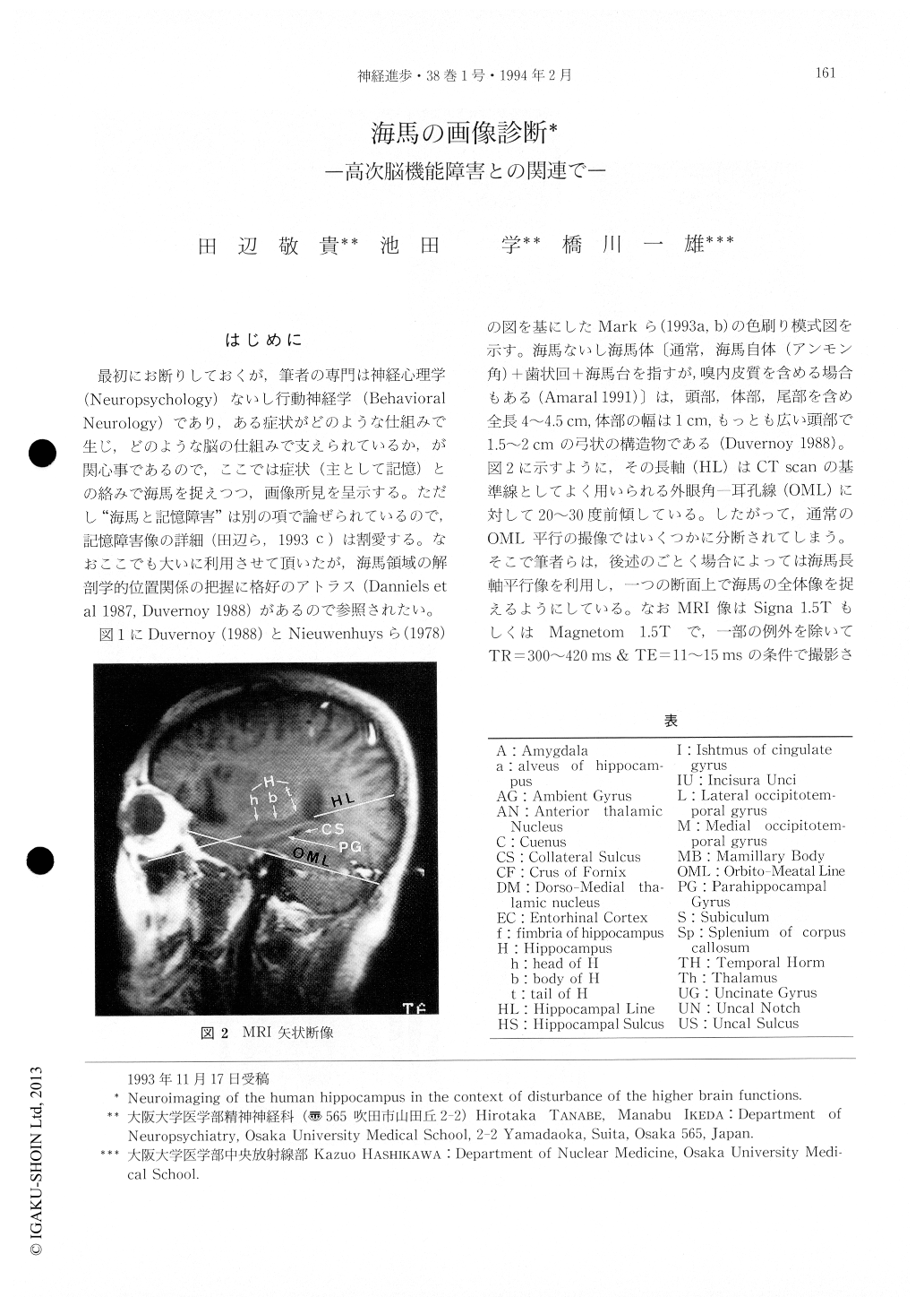
図1にDuvernoy(1988)とNieuwenhuysら(1978)の図を基にしたMarkら(1993a,b)の色刷り模式図を示す。海馬ないし海馬体〔通常,海馬自体(アンモン角)+歯状回+海馬台を指すが,嗅内皮質を含める場合もある(Amaral 1991)〕は,頭部,体部,尾部を含め全長4~4.5cm,体部の幅は1cm,もっとも広い頭部で1.5~2cmの弓状の構造物である(Duvernoy 1988)。図2に示すように,その長軸(HL)はCT scanの基準線としてよく用いられる外眼角―耳孔線(OML)に対して20~30度前傾している。
The hippocampal formation is rather a small structure like a seahorse or a silkworm in shape. Its total length is only between 4 and 4.5 cm, the width of its body on average 1 cm and that of its head 1.5 to 2 cm (Duvernoy). In addition, its long axis is inclined at an angle 20 or 30 degrees negative to the orbitomeatal line (Fig. 2). Accordingly, in images parallel to the orbitomeatal line commonly used, the hippocampal formation is divided into several parts and shown on several separate slices. As a result, it became difficult to evaluate blood flow of the small part in each slice (Fig. 5).

Copyright © 1994, Igaku-Shoin Ltd. All rights reserved.


