Japanese
English
- 有料閲覧
- Abstract 文献概要
- 1ページ目 Look Inside
はじめに
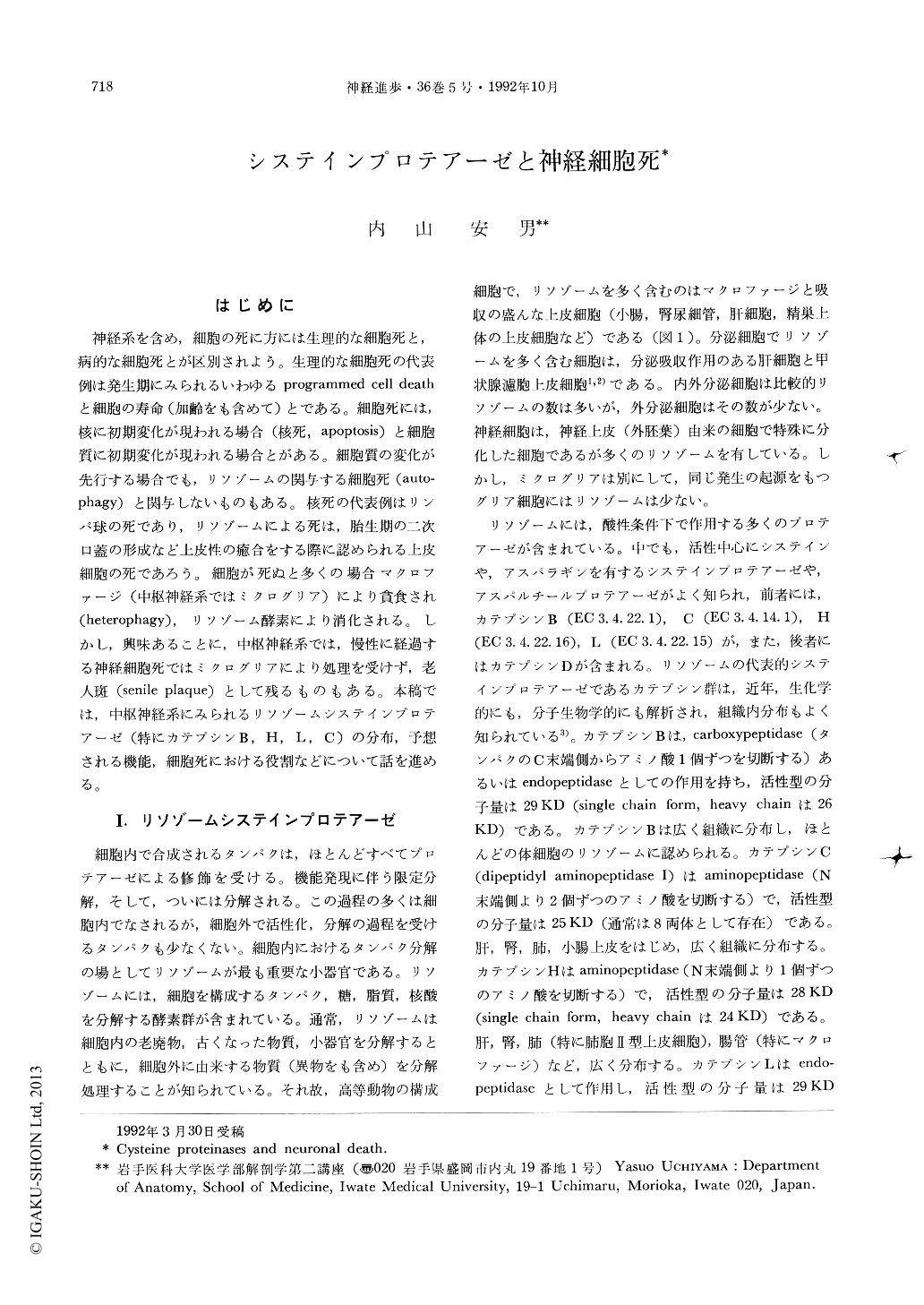
神経系を含め,細胞の死に方には生理的な細胞死と,病的な細胞死とが区別されよう。生理的な細胞死の代表例は発生期にみられるいわゆるprogrammed cell deathと細胞の寿命(加齢をも含めて)とである。細胞死には,核に初期変化が現われる場合(核死,apoptosis)と細胞質に初期変化が現われる場合とがある。細胞質の変化が先行する場合でも,リソゾームの関与する細胞死(autophagy)と関与しないものもある。核死の代表例はリンパ球の死であり,リソゾームによる死は,胎生期の二次口蓋の形成など上皮性の癒合をする際に認められる上皮細胞の死であろう。細胞が死ぬと多くの場合マクロファージ(中枢神経系ではミクログリア)により貧食され(heterophagy),リソゾーム酵素により消化される。しかし,興味あることに,中枢神経系では,慢性に経過する神経細胞死ではミクログリアにより処理を受けず,老人斑(senile plaque)として残るものもある。本稿では,中枢神経系にみられるリソゾームシステインプロテアーゼ(特にカテプシンB,H,L,C)の分布,予想される機能,細胞死における役割などにっいて話を進める。
Lysosomes, a membrane-bound cytoplasmic organelle, contain a great variety of hydrolytic enzymes which are capable of breaking down proteins, nucleic acids, complex carbohydrates and lipids. Cathepsins B, C, H, and L are well characterized cysteine proteinases, each being widely distributed in lysosomes of various mammalian cells. By immunocytochemistry we have shown that these cysteine proteinases are localized not only in lysosomes of various tissue cells but also in secretory granules of certain peptide hormone-producing cells. In pancreatic endocrine B- and A-cells cathepsins B and/or H are localized in crinophagic bodies, which contain dense cores resembling a protein core of secretory granules and are identified as a site to degrade old, unneeded secretory products. Cathepsin B or H is also localized in secretory granules of atrial myoendocrine cells, pituitary pro-opiomelanocortin-pro-ducing cells, and active renin-producing cells including renal juxtaglomerular cells, pituitary LH/FSH cells and submandibular granular ductal cells ; these events suggest that they may participate in the activation processes of peptide hormones.

Copyright © 1992, Igaku-Shoin Ltd. All rights reserved.


