Japanese
English
- 有料閲覧
- Abstract 文献概要
- 1ページ目 Look Inside
I.脊髄空洞症
1.分類
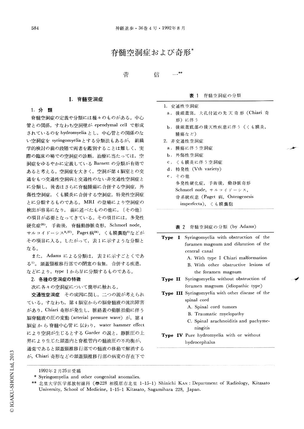
脊髄空洞症の定義や分類には種々のものがある。中心管との関係,すなわち空洞壁がependymal cellで形成されているのをhydromyeliaとし,中心管との関係のない空洞症をsyringomyeliaとする分類法もあるが,組織学的検討の前の段階で両者を鑑別することは難しく,実際の臨床の場での空洞症の診断,治療に当たっては,空洞症をゆるやかに定義しているBarnettの分類が有効であると考える。空洞症を大きく,空洞が第4脳室との交通をもつ交通性空洞病と交通性のない非交通性空洞症とに分類し,後者はさらに脊髄腫瘍に合併する空洞症,外傷性空洞症,くも膜炎に合併する空洞症,特発性空洞症とに分類するものである。MRIの登場により空洞症の検出が容易になり,前に述べたものの他に,〔その他〕の項目が必要となってきている。その項目には,多発性硬化症26),手術後,脊髄動静脈奇形,Schmorl node,サルコイドーシス9,27),Paget病24),くも膜嚢胞8)などがその項目に入る。したがって,表1に示すような分類となる。
また,Adamsによる分類は,表2に示すごとくである1)。頭蓋頸椎移行部での閉塞の有無,合併する疾患,などにより,type IからIVに分類するものである。
The diagnosis of a syringomyelia has been changed dramatically since the introduction of magnet resonance imaging (MRI). The presence of a syrinx cavity is easily demonstrated with MRI. And a follow up study for post operated cases of syringomyelia is well performed with MRI. Syringomyelia is classified into two types, that is, communicating type and non-communication type. The latter is classified further into syringomyelia associated with tumor, arachnoiditis, post traumatic and idiopathic one. Several MR features can diffetentiate each subtype of a syringomyelia. Those findings are septation of a syrinx, CSF flow void sign, high intensity on T2WI, shape of syringobulbia, and Chiari malformation.

Copyright © 1992, Igaku-Shoin Ltd. All rights reserved.


