Japanese
English
- 有料閲覧
- Abstract 文献概要
- 1ページ目 Look Inside
- 参考文献 Reference
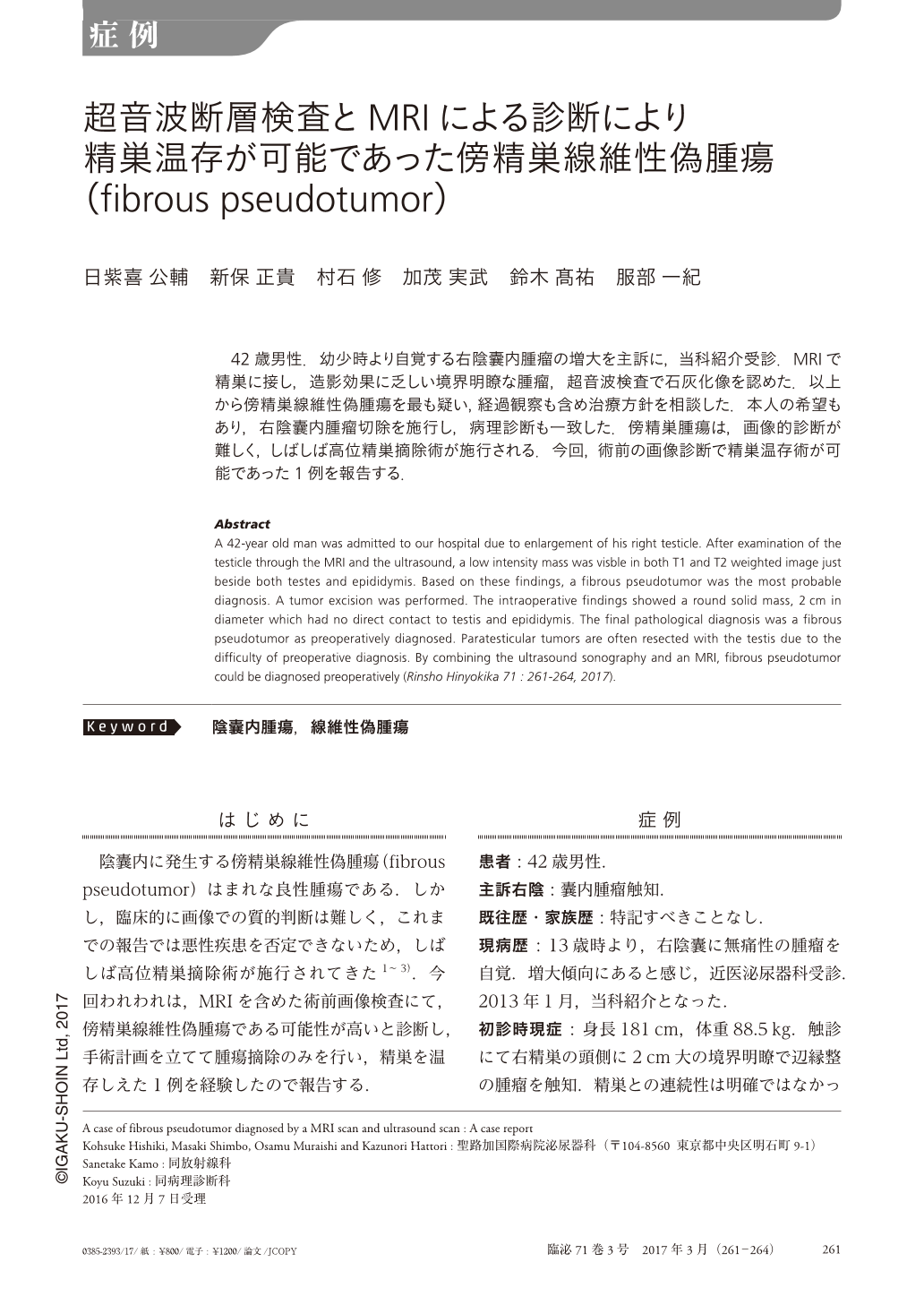
42歳男性.幼少時より自覚する右陰囊内腫瘤の増大を主訴に,当科紹介受診.MRIで精巣に接し,造影効果に乏しい境界明瞭な腫瘤,超音波検査で石灰化像を認めた.以上から傍精巣線維性偽腫瘍を最も疑い,経過観察も含め治療方針を相談した.本人の希望もあり,右陰囊内腫瘤切除を施行し,病理診断も一致した.傍精巣腫瘍は,画像的診断が難しく,しばしば高位精巣摘除術が施行される.今回,術前の画像診断で精巣温存術が可能であった1例を報告する.
Abstract
A 42-year old man was admitted to our hospital due to enlargement of his right testicle. After examination of the testicle through the MRI and the ultrasound, a low intensity mass was visble in both T1 and T2 weighted image just beside both testes and epididymis. Based on these findings, a fibrous pseudotumor was the most probable diagnosis. A tumor excision was performed. The intraoperative findings showed a round solid mass, 2cm in diameter which had no direct contact to testis and epididymis. The final pathological diagnosis was a fibrous pseudotumor as preoperatively diagnosed. Paratesticular tumors are often resected with the testis due to the difficulty of preoperative diagnosis. By combining the ultrasound sonography and an MRI, fibrous pseudotumor could be diagnosed preoperatively (Rinsho Hinyokika 71 : 261-264, 2017).

Copyright © 2017, Igaku-Shoin Ltd. All rights reserved.


