Japanese
English
- 有料閲覧
- Abstract 文献概要
- 1ページ目 Look Inside
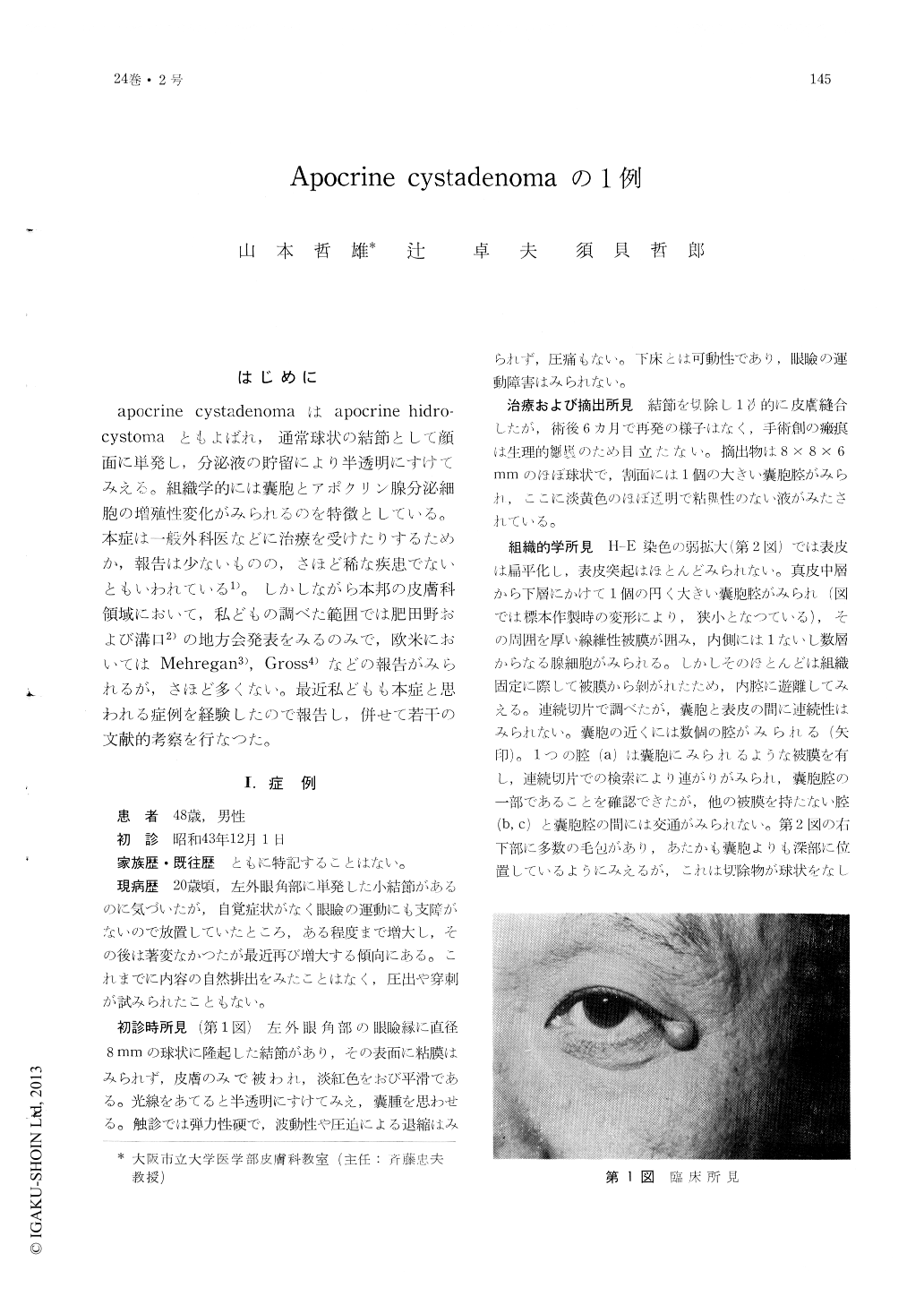
はじめに
apocrine cystadenomaはapocrine hidro-cystomaともよばれ,通常球状の結節として顔面に単発し,分泌液の貯留により半透明にすけてみえる。組織学的には嚢胞とアポクリン腺分泌細胞の増殖性変化がみられるのを特徴としている。本症は一般外科医などに治療を受けたりするためか,報告は少ないものの,さほど稀な疾患でないともいわれている1)。しかしながら本邦の皮膚科領域において,私どもの調べた範囲では肥田野および溝口2)の地方会発表をみるのみで,欧米においてはMehregan3),Gross4)などの報告がみられるが,さほど多くない。最近私どもも本症と思われる症例を経験したので報告し,併せて若干の文献的考察を行なつた。
A case of apocrine cystadenoma in a 48-year-old Japanese man who presented witha slightly reddish, semispherically raised, sessile cystic nodule of 8 mm. in diameter on the left outer canthus. is reported Microscopic studies of sections of the tumor revealed a large cystic space encapsulated with fibrous tissues in the corium. The cyst was lined by one to several layers of secretory epithelium. There were another lumen filled with nests of secretory cells and a dilated secretory duct in the vicinity of the large cyst. The glandular lumen was lined with outer layer of cuboidal immature myoepithelial cells and inner layers of columnar cylind-ric al cells. Enzymatic studies disclosed that the secretory cells were most likely of apocrine origin. The dilated duct was composed of two layers of cuboidal cells, and there was diastase-resistant, PAS-positive secretory debris in the lumen.
Apocrine cystadenoma would not be a retention cyst, but a benign nevoid tumor of apocrine duct origin, because the cystic space was encapsulated with fibrous stroma, and the tumor cells were proliferative and related to the apocrine gland. In view of the fact that the tumor occurred on the canthus, it would be reasonable to consider that it originated from the Moll's gland. To our best knowledge, this is the first confirmed case of apocrine cystadenoma in the Japanese dermatological field.
Copyright © 1970, Igaku-Shoin Ltd. All rights reserved.