Japanese
English
- 有料閲覧
- Abstract 文献概要
- 1ページ目 Look Inside
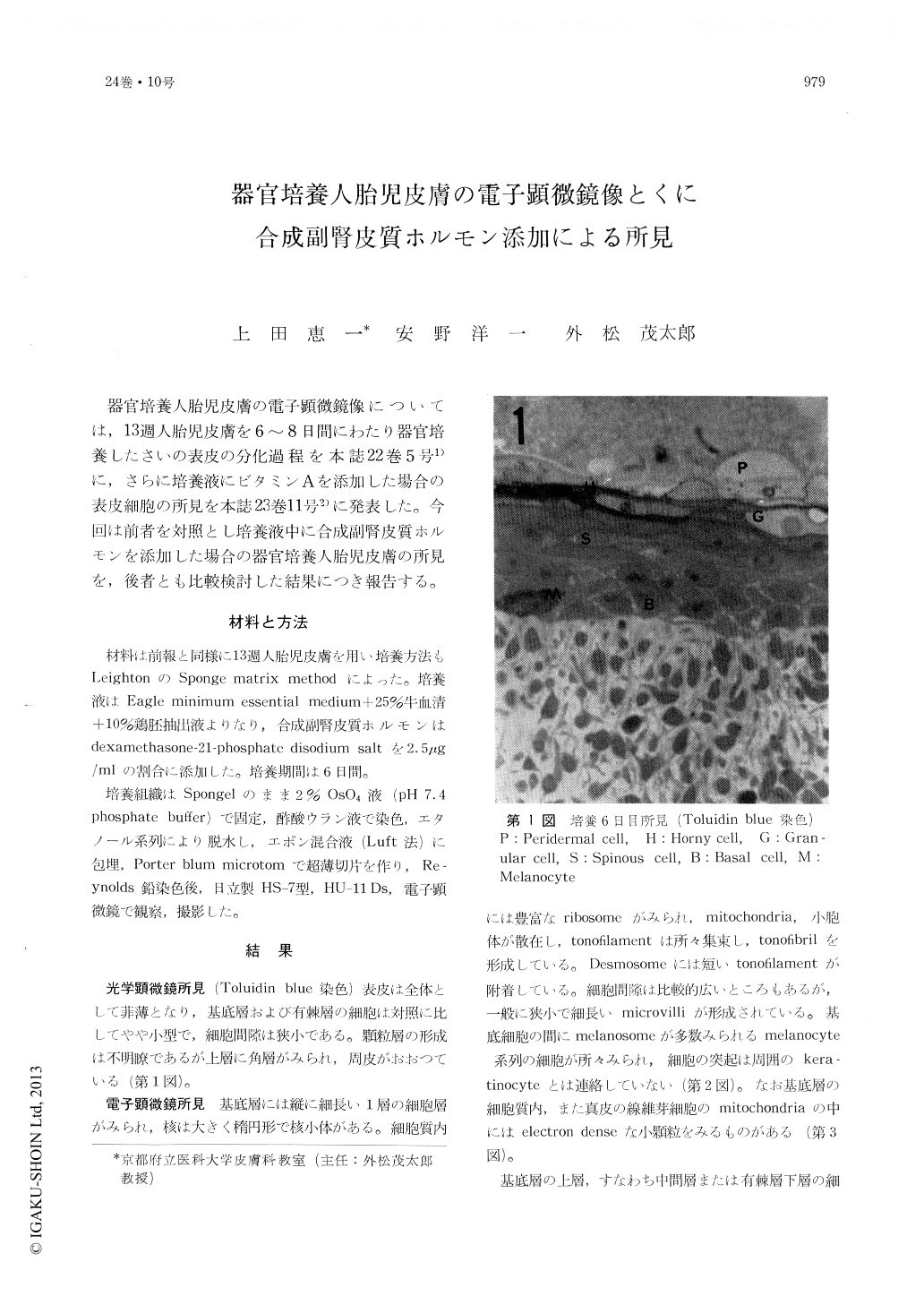
器官培養人胎児皮膚の電子顕微鏡像については,13週人胎児皮膚を6〜8日間にわたり器官培養したさいの表皮の分化過程を本誌22巻5号1)に,さらに培養液にビタミンAを添加した場合の表皮細胞の所見を本誌23巻11号2)に発表した。今回は前者を対照とし培養液中に合成副腎皮質ホルモンを添加した場合の器官培養人胎児皮膚の所見を,後者とも比較検討した結果につき報告する。
The electron microscopic observations were performed on the organ cultured human foetal skin of 13 weeks by the sponge matrix method, especially on the effect of the synthetized adrenocortical hormone. The specimens for EM studies were made from 6 days' culture in the medium containing 2.5 μg of the dexarnethasone-21-phosphate disodium salt in each ml of the Eagle's minimum essential medium.
The control specimens without the hormone were composed of the fully developed epidermis, showing serial changes from the basal layer to the horny layer.
The skin cultured in the medium containing the hormone was thin and the epidermal cells were small as a rule. Although the basal cells showed no significant findings, the intercellular spaces became narrower. The prickle cells formed many microvilli, rather long tonofilaments from the desmosome, compact cytoplasma rich in ribosomes, some of the mitochondria of the cells were long-shaped, but neither the destruction of their membrane nor inhomogeneity of the matrix could be proved. In the upper part of the prickle cell layer there were small keratohyalin granules, the cells in the layer just above which became flat and were filled compactly with fine filaments. Organelles in between them were preserved.
As a conclusion the organ cultured epidermis treated with the steroid hormone showed accela-rated keratinization with the horny layer-like appearance in some parts, well preserved organelles and a suppression of degeneration of them.

Copyright © 1970, Igaku-Shoin Ltd. All rights reserved.


