Japanese
English
- 有料閲覧
- Abstract 文献概要
- 1ページ目 Look Inside
I.緒言
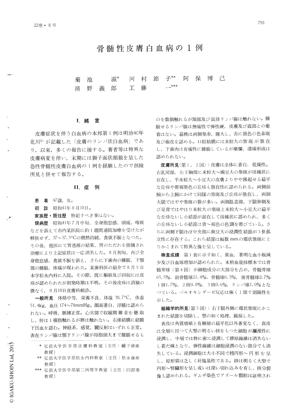
皮膚症状を伴う白血病の本邦第1例は明治40年北川1)が記載した「皮膚のリンパ状白血病」であり,以来,多くの報告に接する。著者等は特異な皮膚病変を伴い,末期には獅子面状顔貌を呈した急性骨髄性皮膚白血病の1例を経験したので剖検所見と併せて報告する。
A 47-year-old woman was admitted to the internal medicine of Hirosaki University Hospital under the diagnosis of myelogenous leukemia on August 18, 1965. At her admission she had marasmus, loss of appetite, gingival swelling and painful swelling of the lower jaw, and the skin manifestations which were composed of purpuric, papular and nodular elements disseminatedor scattered on the chest, upper and lower extremities. Three discrete tumors surrounded by annular purpura were noted on both legs. A few, swollen, thumb-sized submaxillary lymph nodes were found, and the enlarged liver could be palpable one finger-breadth below the costal margin. Gingiva was swollen but did have neither erosion nor ulcer.
Laboratory studies revealed anemia, marked thrombopenia, and leukocytosis, with 65.5 % of myeloblasts in peripheral blood, some of which showed slightly positive peroxydase reaction.
Histologic specimen biopsied from a nodule on the right leg showed a diffuse infiltrate of aty-pical cells with a large pale-staining nucleus and scanty protoplasm throughout the cutis.
While blood transfusion, administration of antibiotics, prednisolone, 6 MP and urethan produced a temporary remission, her condition suddenly turned worse and multiple nodules appeared over the entire body surface, and the patient died on August 30, 1966.
Autopsy revealed nodules and tumors over the entire body which predominated on the face, root of the tongue, tonsils, epiglottis and submandibular region. They pressed the pharyngeal region. Many lymph nodes along the esophagus and respiratory tract and in the abdominal cavity were enlarged. Histologic specimen showed infiltration of leukemic cells in all organs of the body.

Copyright © 1968, Igaku-Shoin Ltd. All rights reserved.


