Japanese
English
- 有料閲覧
- Abstract 文献概要
- 1ページ目 Look Inside
はじめに
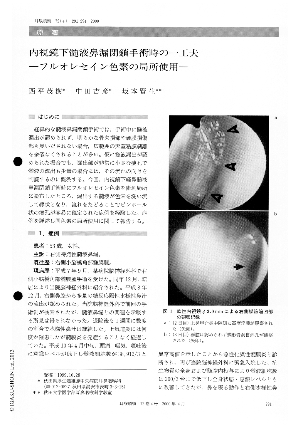
経鼻的な髄液鼻漏閉鎖手術では,手術中に髄液漏出が認められず,明らかな骨欠損部や硬膜損傷部も見いだされない場合,広範囲の天蓋粘膜剥離を余儀なくされることが多い。仮に髄液漏出が認められた場合でも,漏出部が非常に小さな瘻孔で髄液の流出も少量の場合には,その流れの向きを判読するのに難渋する。今回,内視鏡下経鼻髄液鼻漏閉鎖手術時にフルオレセイン色素を術創局所に塗布したところ,漏出する髄液が色素を洗い流して線状となり,流れをたどることでピンホール状の瘻孔が容易に確定された症例を経験した。症例を詳述し同色素の局所使用に関して報告する。
A 53-year-old female was hospitalized because of meningitis on April 1998. The patient received an operation due to CP angle meningioma in 1995. and the patient complained of persistent CSF rhinorrhea since December, 1996. Although neurosurgeonsexamined the causative connection between the CSF rhinorrhea and the prerivous operation, no pathological findings had been demonstrated.
Nasal endoscopic examination with the excellent visualization of the telescope combining with topi-cal use of fluorescein dye was very helpful to iden-tify and localize a pin hole fistula at the right side cribriform plate. Leakage was successfully stopped by the fascia lata grafting with fibrin glue. No recurrence of CSF rhinorrhea or meningitis have been occurred for 12 months.

Copyright © 2000, Igaku-Shoin Ltd. All rights reserved.


