Japanese
English
原著
診断困難であった鼻性眼窩内骨膜下嚢胞の1症例
Intraorbital Subperiosteal Pyocele as a Complication of Sinusitis:Report of a Case
水谷 陽江
1
,
伊藤 光子
2
,
金子 行子
3
,
石井 哲夫
4
Kiyoe Mizutani
1
1東京女子医科大学耳鼻咽喉科教室
2至誠会第二病院耳鼻咽喉科
3至誠会第二病院眼科
4東京女子医科大学耳鼻咽喉科教室
1Department of Otorhinolaryngology, Tokyo Women's Medical College
pp.375-378
発行日 1992年5月20日
Published Date 1992/5/20
DOI https://doi.org/10.11477/mf.1411900539
- 有料閲覧
- Abstract 文献概要
- 1ページ目 Look Inside
はじめに
副鼻腔と眼窩は隣接臓器であるために,副鼻腔疾患により種々の眼症状をきたすことが多く,眼球突出,視力障害などはしばしば経験する。
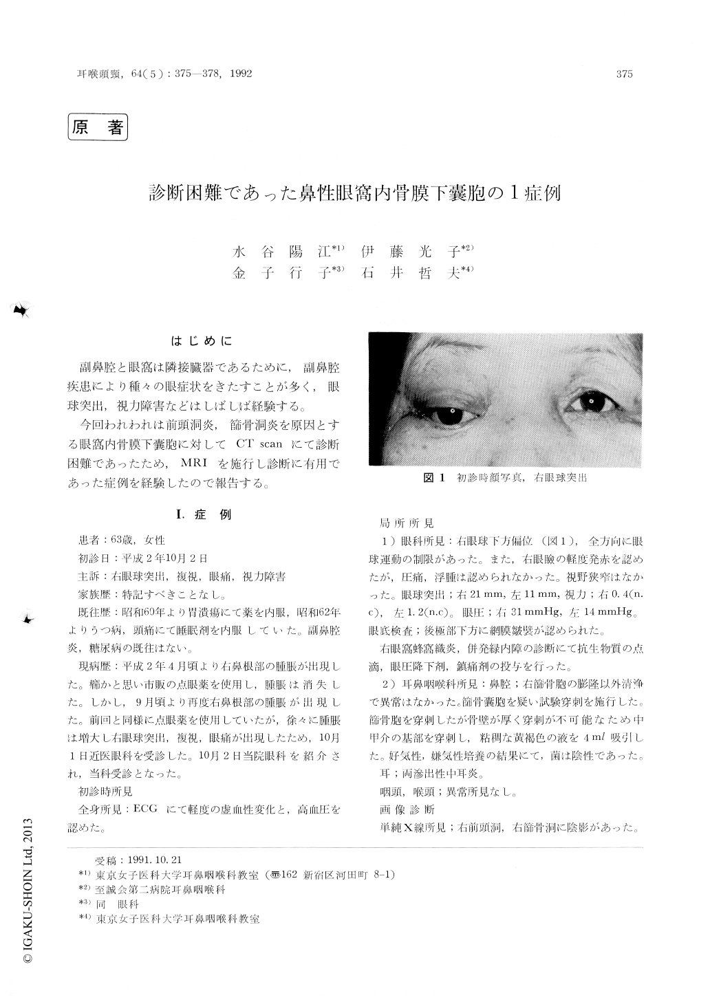
今回われわれは前頭洞炎,篩骨洞炎を原因とする眼窩内骨膜下嚢胞に対してCT scanにて診断困難であったため,MRIを施行し診断に有用であった症例を経験したので報告する。
A 63-year-old woman with an intraorbital subperiosteal pyocele was presented. Intraorbital subperiosteal pyocele occurred secondary to fron-tal and ethmoidal sinusitis. Relationship between them was not clear by CT scan. However, MRI revealed the intraorbital subperiosteal pyocele as a complication of frontal and ethmoidal sinusitis.
MRI is an useful examination for the diagnosis of the soft tissue masses.

Copyright © 1992, Igaku-Shoin Ltd. All rights reserved.


