Japanese
English
- 有料閲覧
- Abstract 文献概要
- 1ページ目 Look Inside
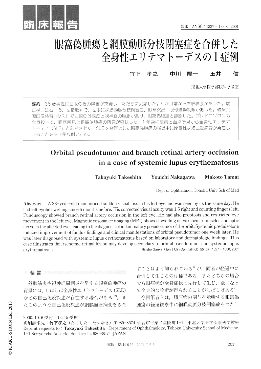
36歳男性に左眼の視力障害が突発し,ただちに受診した。6か月前から左眼腫脹があった。矯正視力は右1.5,左指数弁で,左眼に網膜動脈分枝閉塞症,眼球突出,眼球運動制限があった。磁気共鳴画像検査(MRI)で左眼の外眼筋と視神経の腫脹があり,眼窩偽腫瘍と診断した。プレドニゾロンの全身投与で,眼底所見と眼窩偽腫瘍の所見が軽快した。1年後に皮膚と血液所見から全身性エリテマトーデス(SLE)と診断された。SLEを背景とした眼窩偽腫瘍の経過中に閉塞性網膜血管病変が発症しうることを示す稀な例である。
A 36-year-old man noticed sudden visual loss in his left eye and was seen by us the same day. He had left eyelid swelling since 6 months before. His corrected visual acuity was 1.5 right and counting fingers left. Funduscopy showed branch retinal artery occlusion in the left eye. He had also proptosis and restricted eye movement in the left eye. Magnetic resonance imaging (MRI) showed swelling of extraocular muscles and optic nerve in the affected eye, leading to the diagnosis of inflammatory pseudotumor of the orbit. Systemic prednisolone induced improvement of fundus findings and clinical manifestations of orbital pseudotumor one week later. He was later diagnosed with systemic lupus erythematosus based on laboratory and dermatologic findings. This case illustrates that ischemic retinal lesion may develop secondary to orbital pseudotumor and systemic lupus erythematosus.

Copyright © 2001, Igaku-Shoin Ltd. All rights reserved.


