Japanese
English
- 有料閲覧
- Abstract 文献概要
- 1ページ目 Look Inside
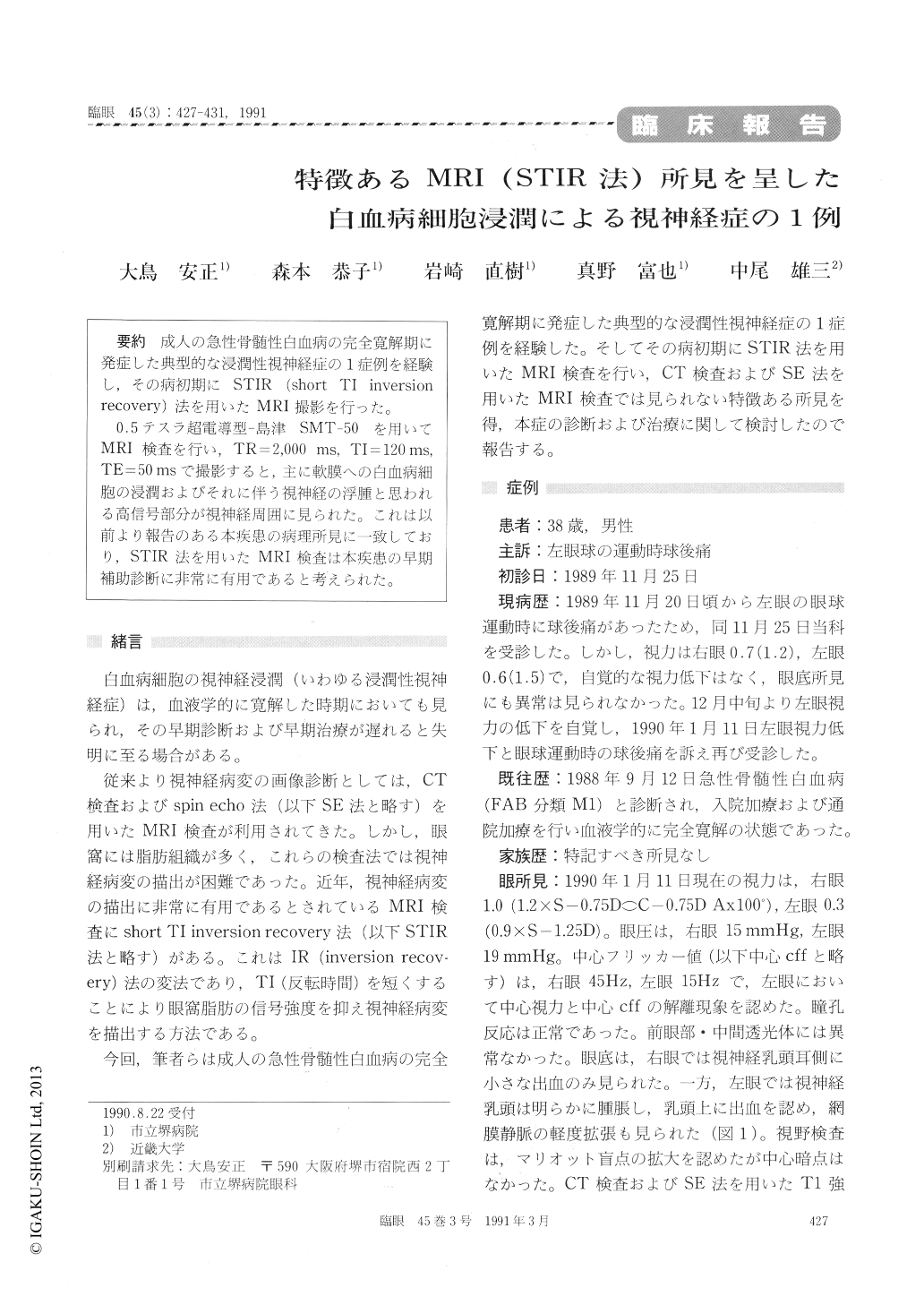
成人の急性骨髄性白血病の完全寛解期に発症した典型的な浸潤性視神経症の1症例を経験し,その病初期にSTIR (short TI inversionrecovery)法を用いたMRI撮影を行った。
0.5テスラ超電導型—島津 SMT-50 を用いてMRI検査を行い,TR=2,000ms,TI=120ms,TE=50msで撮影すると,主に軟膜への白血病細胞の浸潤およびそれに伴う視神経の浮腫と思われる高信号部分が視神経周囲に見られた。これは以前より報告のある本疾患の病理所見に一致しており,STIR法を用いたMRI検査は本疾患の早期補助診断に非常に有用であると考えられた。
A 38-year-old male presented with pain in his left eye during eye movement. He had been diagnosed as acute myelocytic leukemia 14 months before. He was in a state of clinical cure when seen by us. We detected optic disc edema in the left eye. Magnetic resonance imaging (MRI) using short TI inversion recovery (STIR) technique revealed a high-signal lesion around the left optic nerve sug-gesting infiltration by leukemic cells. Optic disc edema disappeared 10 weeks after Linac irradiation totalling 26 Gy. MRI with STIR technique was thus instrumental in the diagnosis of leukemic infiltra-tion of the optic nerve in this patient.

Copyright © 1991, Igaku-Shoin Ltd. All rights reserved.


