Japanese
English
- 有料閲覧
- Abstract 文献概要
- 1ページ目 Look Inside
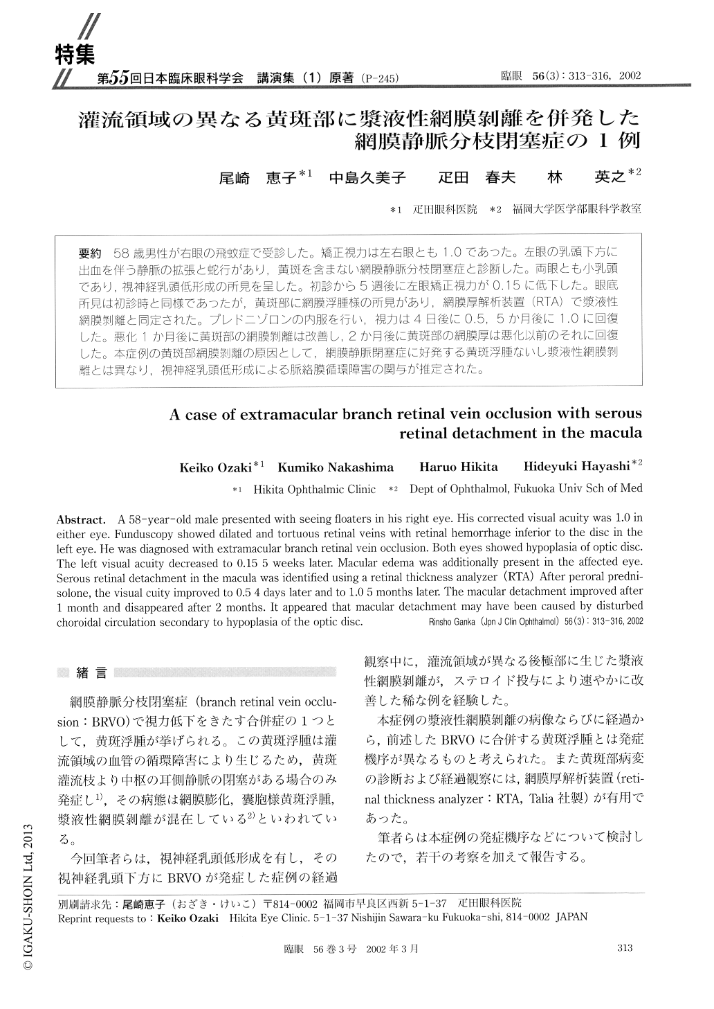
(P−245) 58歳男性が右眼の飛蚊症で受診した。矯正視力は左右眼とも1.0であった。左眼の乳頭下方に出血を伴う静脈の拡張と蛇行があり,黄斑を含まない網膜静脈分枝閉塞症と診断した。両眼とも小乳頭であり,視神経乳頭低形成の所見を呈した。初診から5週後に左眼矯正視力が0.15に低下した。眼底所見は初診時と同様であったが,黄斑部に網膜浮腫様の所見があり,網膜厚解析装置(RTA)で漿液性網膜剥離と同定された。プレドニゾロンの内服を行い,視力は4日後に0.5,5か月後に1.0に回復した。悪化1か月後に黄斑部の網膜剥離は改善し,2か月後に黄斑部の網膜厚は悪化以前のそれに回復した。本症例の黄斑部網膜剥離の原因として,網膜静脈閉塞症に好発する黄斑浮腫ないし漿液性網膜剥離とは異なり,視神経乳頭低形成による脈絡膜循環障害の関与が推定された。
A 58-year-old male presented with seeing floaters in his right eye. His corrected visual acuity was 1.0 in either eye. Funduscopy showed dilated and tortuous retinal veins with retinal hemorrhage inferior to the disc in the left eye. He was diagnosed with extramacular branch retinal vein occlusion. Both eyes showed hypoplasia of optic disc. The left visual acuity decreased to 0.15 5 weeks later. Macular edema was additionally present in the affected eye. Serous retinal detachment in the macula was identified using a retinal thickness analyzer (RTA) After peroral predni-solone, the visual cuity improved to 0.5 4 days later and to 1.0 5 months later. The macular detachment improved after 1 month and disappeared after 2 months. It appeared that macular detachment may have been caused by disturbed choroidal circulation secondary to hypoplasia of the optic disc.

Copyright © 2002, Igaku-Shoin Ltd. All rights reserved.


