Japanese
English
- 有料閲覧
- Abstract 文献概要
- 1ページ目 Look Inside
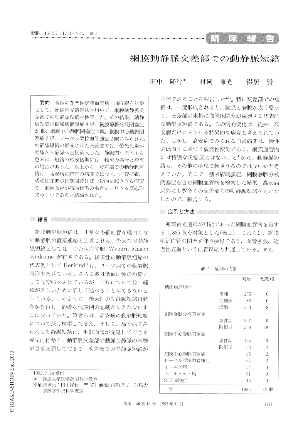
各種の閉塞性網膜血管病1,885眼を対象として,連続螢光造影法を用いて,網膜動静脈交差部での動静脈短絡を検索した。その結果,動静脈短絡は糖尿病網膜症8眼,網膜静脈分枝閉塞症28眼,網膜中心静脈閉塞症2眼,網膜中心動脈閉塞症2眼,レーベル粟粒血管腫症2眼にみられた。動静脈短絡の形成された交差部では,螢光色素が動脈から静脈へ直接流入した。静脈内へ流入する色素は,短絡の形成初期には,軸流の場合と層流の場合があった。以上から,交差部での動静脈短絡は,高安病に特有の病変ではなく,血管拡張,透過性亢進が長期間続けば一般的に起きうる病変で,網膜血管が病的状態の場合にとりうる反応形式の1つであると結論された。
We reviewed fluorescein angiograms in 1,885 eyes with various occlusive retinal vascular dis-eases to detect arteriovenous shunt at arter-iovenous crossings. We could identify arteriovenous shunt in diabetic retinopathy 8 eyes, branch retinal vein occlusion 28 eyes, central retinal vein occlu-sion 2 eyes and miliary angiomatosis of Leber 2 eyes. Fluorescent dye flowed from artery into vein at the site of shunting manifesting either axial flow or laminar flow pattern. The findings seemed to indicate that shunting at arteriovenous crossings may occur in various retinal vascular diseases other than pulseless disease. We postulate that arter- iovenous shunting is one of the basic patterns of vascular reaction in the retina in the presence of persistent vasodilatation and increased permeabil-ity.

Copyright © 1992, Igaku-Shoin Ltd. All rights reserved.


