Japanese
English
- 有料閲覧
- Abstract 文献概要
- 1ページ目 Look Inside
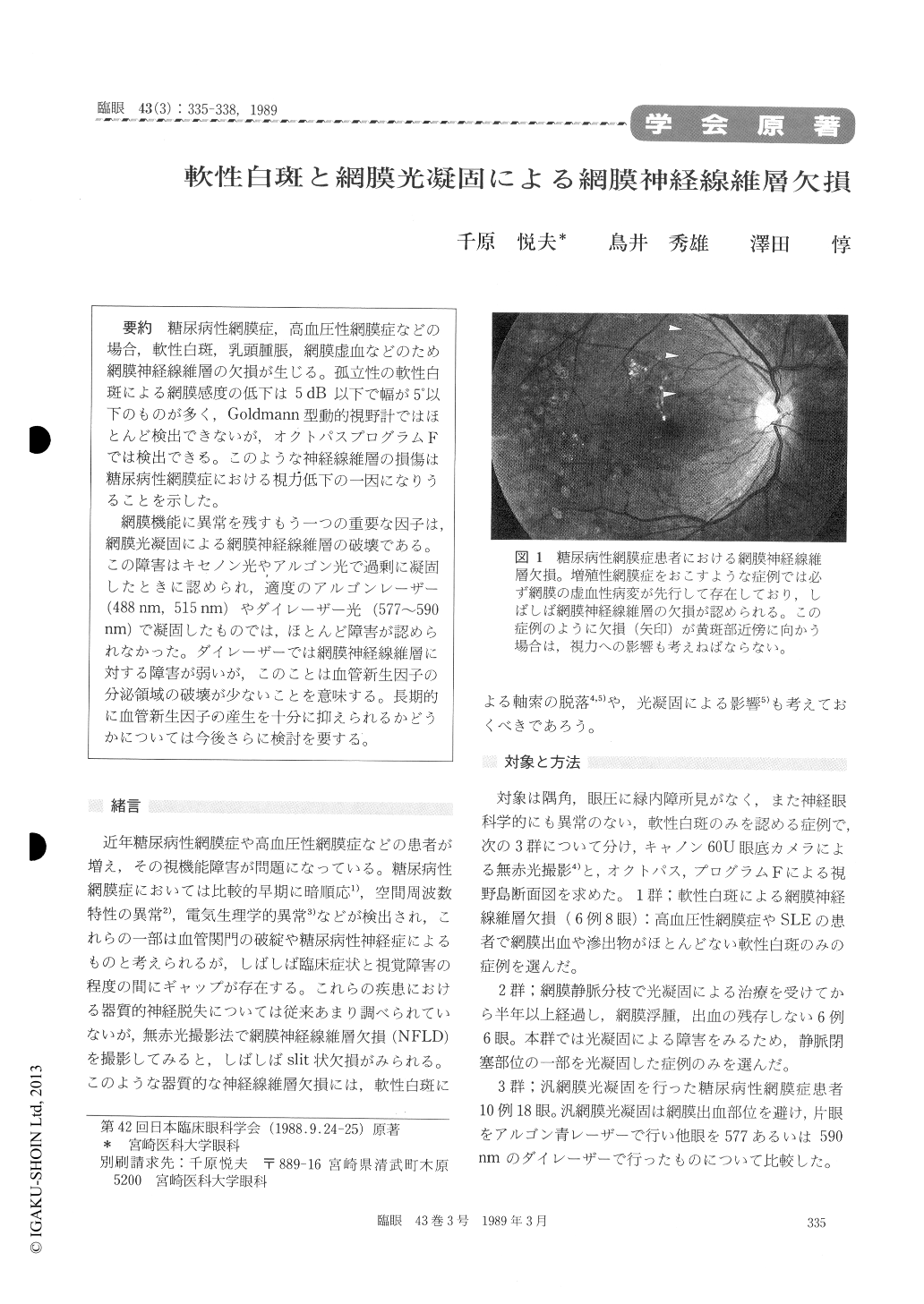
糖尿病性網膜症,高血圧性網膜症などの場合,軟性白斑,乳頭腫脹,網膜虚血などのため網膜神経線維層の欠損が生じる。孤立性の軟性白斑による網膜感度の低下は5dB以下で幅が5°以下のものが多く,Goldmann型動的視野計ではほとんど検出できないが,オクトパスプログラムFでは検出できる。このような神経線維層の損傷は糖尿病性網膜症における視力低下の一因になりうることを示した。
網膜機能に異常を残すもう一つの重要な因子は,網膜光凝固による網膜神経線維層の破壊である。この障害はキセノン光やアルゴン光で過剰に凝固したときに認められ,適度のアルゴンレーザー(488nm,515nm)やダイレーザー光(577〜590nm)で凝固したものでは,ほとんど障害が認められなかった。ダイレーザーでは網膜神経線維層に対する障害が弱いが,このことは血管新生因子の分泌領域の破壊が少ないことを意味する。長期的に血管新生因子の産生を十分に抑えられるかどうかについては今後さらに検討を要する。
We evaluated retinal nerve fiber layer defects (NFLD) in 8 eyes with cotton-wool spots, in 6 eyes with branch retinal vein occlusion treated by photocoagulation, and in 18 eyes with diabetic retinopathy treated by panretinal photocoagula-tion. Either xenon, argon laser or dye laser (577 to 590 nm in wavelength) was used for retinal photocoagulation. The eyes were studied by static perimetry and by fundus photography using red -free light.
NFLDs secondary to solitary cotton-wool spots were small, with threshold changes less than 5dB in depth and 5 degrees in width. These changes could be detected by program F of Octopus perimeter and not by Goldmann kinetic perimeter. The NFLDs caused visual dysfunctions in eyes with diabetic retinopathy.
NFLDs developed after excessive photocoagula-tion with xenon or argon laser. Adequate coagula-tion with dye laser or argon blue laser caused very mild NFLDs. Such weak damages to the nerve fiber layer appeared to be ineffective to suppress vasoformative factor from the neurosensory retina.

Copyright © 1989, Igaku-Shoin Ltd. All rights reserved.


