Japanese
English
- 有料閲覧
- Abstract 文献概要
- 1ページ目 Look Inside
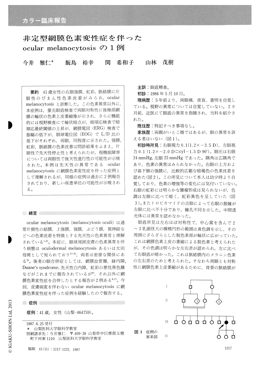
41歳女性の右眼強膜,虹彩,脈絡膜に片眼性のびまん性色素沈着がみられ,ocularmelanocytosisと診断した。この色素異常以外に,本症例は,螢光眼底検査で両眼対称性に後極部網膜の輪状の色素上皮萎縮像が示され,さらに機能的には視野検査にて輪状暗点が,暗順応検査で暗順応最終閾値の上昇が,網膜電図(ERG)検査で振幅の低下が,眼球電位図(EOG)でL/D比の低下がそれぞれ,両眼,同程度に示された。強膜,虹彩,脈絡膜の色素沈着は問診結果をふまえ,片眼性で先天性停止性と考えられたが,視機能障害については両眼性で後天性進行性の可能性が示唆された。本例は先天性の異常であるocularmelanocytosisに網膜色素変性症を伴った症例として理解されるが,同様の症例は過去に2例報告されており,新しい疾患単位の可能性が示唆される。
A 41-year-old female presented with hyperpig-mentation of the sclera, iris and choroid in her right eye. The condition was present since childhood and was diagnosed as ocular melanocytosis. Fun-duscopy revealed abnormal pigmentation in both fundi. When seen by fluorescein angiography, the retinal pigment epithelium was atrophic in the macular area. The visual acuity and color vision were normal. Visual field testing showed symmetri-cal paracentral ring scotomas. Adaptometry showed normal threshold for cone and elevated threshold for rod. Electroretinography showed diminished amplitudes of scotopic, photopic andbright flash ERU5. Electrooculogram was subnor-mal with low L/D ratio. Although the fundus appearance lacked the char-acteristic findings of attenuated retinal arteries and bone-spicule black pigments, the functional changes were compatible to those of retinitis pig-mentosa.
There are two reported cases of oculodermal melanocytosis associated with retinitis pigmentosa. ' In view of the close relation between ocular and oculodermal melanocytosis the associated occur-rence of retinitis pigmentosa and ocular or oculodermal melanocytosis might constitute a new syndrome.
Rinsho Ganka (Jpn J Clin Ophthalmol) 41(11) : 1217-1222,1987

Copyright © 1987, Igaku-Shoin Ltd. All rights reserved.


