Japanese
English
- 有料閲覧
- Abstract 文献概要
- 1ページ目 Look Inside
二次的人工水晶体移植術を行った94例104眼について,術前,術後の角膜内皮細胞をSpecular microscopeを用いて観察した.
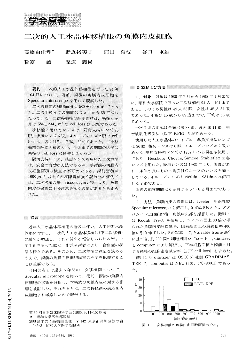
二次移植前の細胞面積は502±240μm2であった.二次手術までの期間は2カ月から35年にわたっていた.二次移植後の細胞面積は,術後6ヵ月で584±234μm2でcell lossは14%であった.二次移植に用いたレンズは,隅角支持レンズ96眼,後房レンズ6眼,4ループレンズ2眼でcelllossは,各々11%,7%,22%であった.二次移植前の細胞面積の大小,手術までの期間の因子は,術後のcell lossに影響しなかった.
隅角支持レンズ,後房レンズを用いた二次移植は,安全で有効な方法であるが,手術前の角膜内皮細胞面積の検査は不可欠である.術前面積が1000μm2以上で内皮障害が強く疑われる症例では,二次移植の際,viscosurgery等により,角膜内皮の保護に十分注意を払う必要があると考えられた.
We evaluated the state of corneal endothelium before and after secondary intraocularlens implantation in 104 cases with the use of specular microscopy. The interval between the initial cataract extraction and lens im-plantation ranged from 2 months to 35 years, average 5.5 years.
The cell area of corneal endothelium before lens implantation averaged 502.4±246 μm2. After surgery, there was a mean loss of the corneal endothelial cells by 14%. The rate of loss differed depending on the type of the intraocular lens : 11% for angle-support lens (96 eyes), 7% for posterior chamber lens (6 eyes) and 22% for iris clip lens (2 eyes).
There were 4 eyes in which the endothelial cell area was greater than 1000 /./ mz before surgery and which intraocular lens implantation was a success. Examina-tion of the corneal endothelium is recommended as a routine procedure prior to secondary lens implantation in order to detect cellular abnormalities and to take appropriate measures during surgery.
Copyright © 1986, Igaku-Shoin Ltd. All rights reserved.