Japanese
English
臨床報告
Iridocorneal endothelial syndrome5例におけるスペキュラーマイクロスコープによる観察
Specular microscopy in five cases with iridocorneal endothelial syndrome
原田 敬志
1
,
三浦 元也
2
,
田辺 吉彦
3
,
後藤 修
4
Takashi Harada
1
,
Motoya Miura
2
,
Yoshihiko Tanabe
3
,
Osamu Goto
4
1名大分院眼科
2名大眼科
3中部労災病院眼科
4岡崎市民病院眼科
1Br. Hosp. Service of Ophthalmol., Nagoya Univ. Sch. of Med.
2Dept. of Ophthalmol., Sch. of Med., Nagoya Univ.
3Service of Ophthalmol.,Chubu-Rousai Hosp.
4Service of Ophthalmol., Okazaki Municip. Hosp.
pp.1363-1367
発行日 1985年12月15日
Published Date 1985/12/15
DOI https://doi.org/10.11477/mf.1410209577
- 有料閲覧
- Abstract 文献概要
- 1ページ目 Look Inside
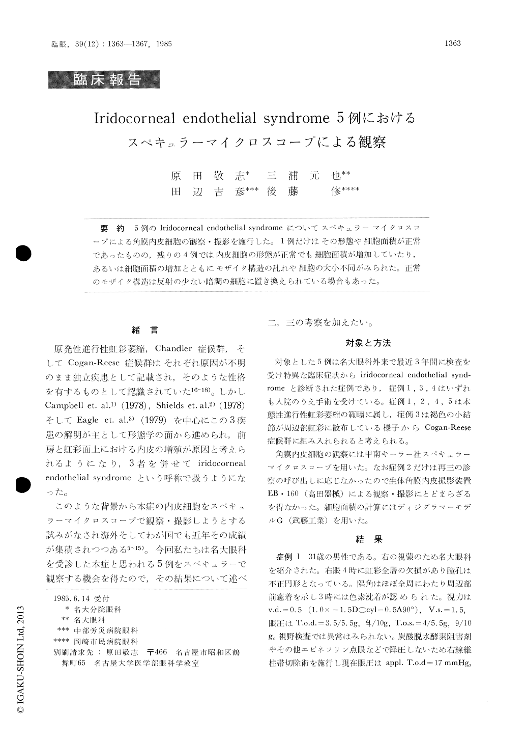
5例のIridocorneal endothelial syndromeについてスペキュラーマイクロスコープによる角膜内皮細胞の観察・撮影を施行した.1例だけはその形態や細胞面積が正常であったものの,残りの4例では内皮細胞の形態が正常でも細胞面積が増加していたり,あるいは細胞面積の増加とともにモザイク構造の乱れや細胞の大小不同がみられた.正常のモザイク構造は反射の少ない暗調の細胞に置き換えられている場合もあった.
We evaluated the corneal endothelium in 5 cases with iridocorneal endothelial syndrome with the use of specular microscopy. Only one case manife-sted normal corneal endothelium. In the other 4 eyes, the corneal endothelium showed pleomorphism and blurring of the endothelial cell margin. Discrete black-out areas were another characteristic feature resulting from replacement of some normal endo-thelial cells by pathologic, dark-appearing ones.

Copyright © 1985, Igaku-Shoin Ltd. All rights reserved.


