Japanese
English
- 有料閲覧
- Abstract 文献概要
- 1ページ目 Look Inside
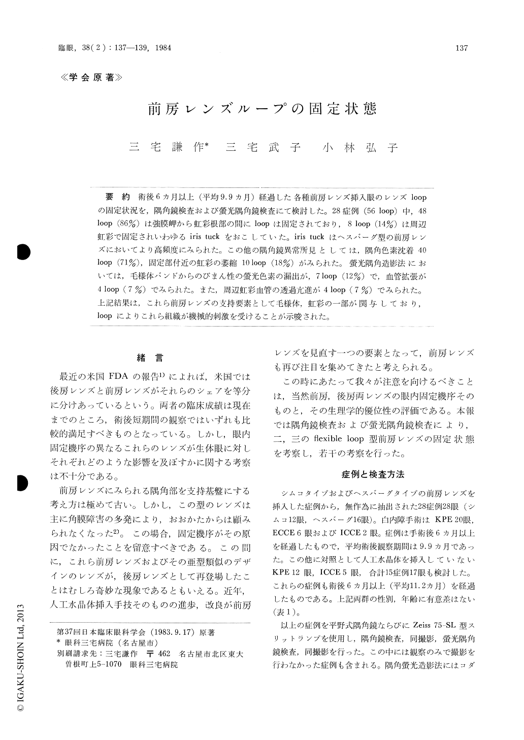
術後6ヵ月以上(平均9.9ヵ月)経過した各種前房レンズ挿入眼のレンズloopの固定状況を,隅角鏡検査および螢光隅角鏡検査にて検討した。28症例(56 loop)中,48loop (86%)は強膜岬から虹彩根部の間にIoopは固定されており,8 loop (14%)は周辺虹彩で固定されいわゆるiris tuckをおこしていた。iris tuckはヘスバーグ型の前房レンズにおいてより高頻度にみられた。この他の隅角鏡異常所見としては,隅角色素沈着40loop (71%),固定部付近の虹彩の萎縮10 loop (18%)がみられた。螢光隅角造影法においては,毛様体バンドからのびまん性の螢光色素の漏出が,7 loop (12%)で,血管拡張が4 loop (7%)でみられた。また,周辺虹彩血管の透過亢進が4 loop (7%)でみられた。上記結果は,これら前房レンズの支持要素として毛様体,虹彩の一部が関与しており,loopによりこれら組織が機械的刺激を受けることが示唆された。
Postoperative gonioscopic and fluorescein gonio-scopic examinations were carried out in 28 cases that underwent anterior chamber lens implanta-tion with Simco and Hesburg type lenses. The examinations were performed at least 6 months after implantation (average 9.9 months). Of the 56 loops examined, 48 were placed between the scleral spur and the iris root in contact with the ciliary band, and the other 8 were placed peripheral to the iris, resulting in iris tuck. Gonioscopy revealed pig-mentation at the trabecular meshwork in 40 loops and atrophy of the iris in the vicinity of the loop in 10 loops.

Copyright © 1984, Igaku-Shoin Ltd. All rights reserved.


