Japanese
English
- 有料閲覧
- Abstract 文献概要
- 1ページ目 Look Inside
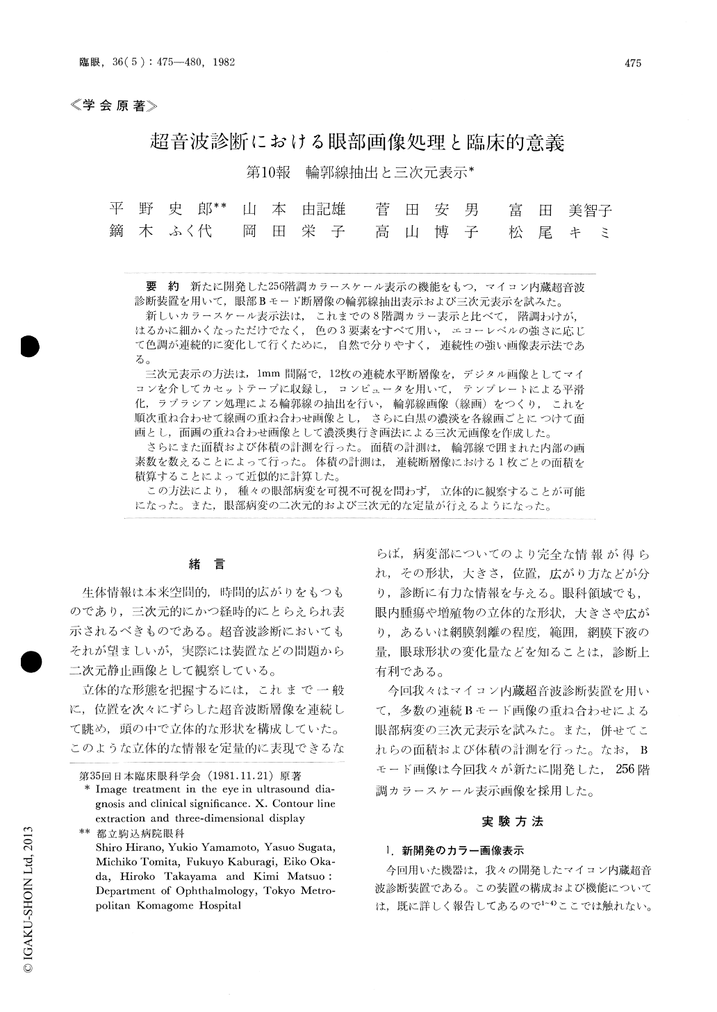
新たに開発した256階調カラースケール表示の機能をもつ,マイコン内蔵超音波診断装置を用いて,眼部Bモード断層像の輪郭線抽出表示および三次元表示を試みた。
新しいカラースケール表示法は,これまでの8階調カラー表示と比べて,階調わけが,はるかに細かくなっただけでなく,色の3要素をすべて用い,エコーレベルの強さに応じて色調が連続的に変化して行くために,自然で分りやすく,連続性の強い画像表示法である。
三次元表示の方法は,1mm間隔で,12枚の連続水平断層像を,デジタル画像としてマイコンを介してカセットテープに収録し,コンピュータを用いて,テンプレートによる平滑化,ラプラシアン処理による輪郭線の抽出を行い,輪郭線画像(線画)をつくり,これを順次重ね合わせて線画の重ね合わせ画像とし,さらに白黒の濃淡を各線画ごとにつけて面画とし,面画の重ね合わせ画像として濃淡奥行き画法による三次元画像を作成した。
さらにまた面積および体積の計測を行った。面積の計測は,輪郭線で囲まれた内部の画素数を数えることによって行った。体積の計測は,連続断層像における1枚ごとの面積を積算することによって近似的に計算した。
この方法により,種々の眼部病変を可視不可視を問わず,立体的に観察することが可能になった。また,眼部病変の二次元的および三次元的な定量が行えるようになった。
This report describes two methods of three-dimensional display of intraocular tissues obtained from a series of consecutive cross sectional (B-mode) images.
One is a line image consisting of a set of overlaid contour lines. The other is a shaded image consist-ing of a set of gradually darkened planes which is also obtained from the corresponding B-scan digi-tized images by using image processing techniques such as smoothing and Laplacian with the aid of microcomputer. The appearance of the tissue may be observed at any visual angle. Further, we can make a measurement of areas and volumes of the tissue by this system.

Copyright © 1982, Igaku-Shoin Ltd. All rights reserved.


