Japanese
English
- 有料閲覧
- Abstract 文献概要
- 1ページ目 Look Inside
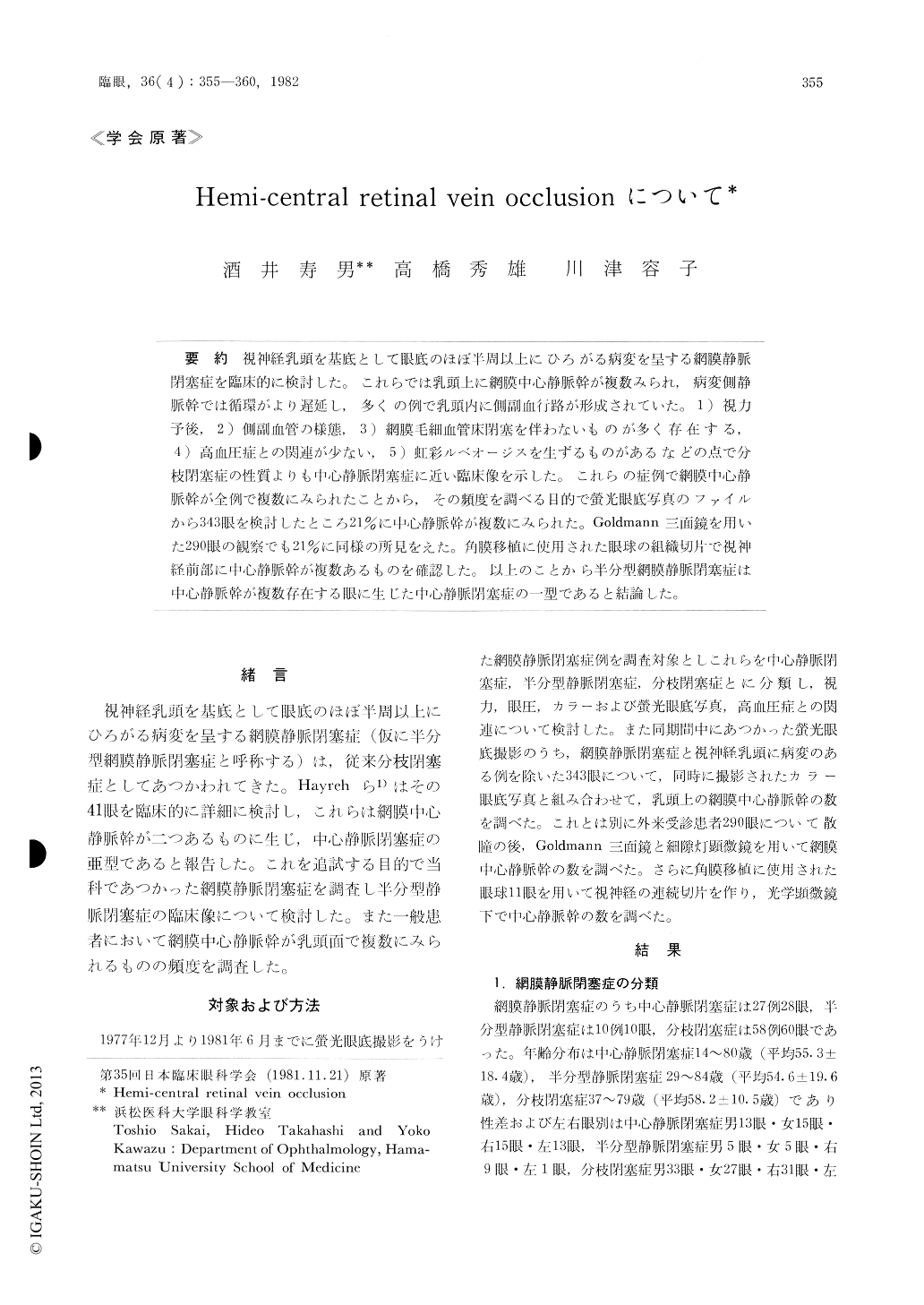
視神経乳頭を基底として眼底のほぼ半周以上にひろがる病変を呈する網膜静脈閉塞症を臨床的に検討した。これらでは乳頭上に網膜中心静脈幹が複数みられ,病変側静脈幹では循環がより遅延し,多くの例で乳頭内に側副血行路が形成されていた。1)視力予後,2)側副血管の様態,3)網膜毛細血管床閉塞を伴わないものが多く存在する,4)高血圧症との関連が少ない,5)虹彩ルベオージスを生ずるものがあるなどの点で分枝閉塞症の性質よりも中心静脈閉塞症に近い臨床像を示した。これらの症例で網膜中心静脈幹が企例で複数にみられたことから,その頻度を調べる目的で螢光眼底写真のファイルから343限を検討したところ21%に中心静脈幹が複数にみられた。Goldmann三面鏡を用いた290眼の観察でも21%に同様の所見をえた。角膜移植に使用された眼球の組織切片で視神経前部に中心静脈幹が複数あるものを確認した。以上のことから半分型網膜静脈閉塞症は中心静脈幹が複数存在する眼に生じた中心静脈閉塞症の一型であると結論した。
We evaluated 10 cases of hemi-central retinal vein occlusion (hemi-CRVO, Hayreh 1980) com-paring its clinical features with those of CRVO and BRVO. In hemi-CRVO, the condition appeared as venous stasis retinopathy and was associated with formation of collateral vessels in the disc in 7 cases each. One case developed rubeosis iridis with ele-vated intraocular pressure. Systemic hypertension was present in 4 and was absent in 6 cases. These features simulated CRVO rather than BRVO. We counted the number of central retinal veins withinthe disc in our 10 cases and in 633 control eyes.

Copyright © 1982, Igaku-Shoin Ltd. All rights reserved.


