Japanese
English
- 有料閲覧
- Abstract 文献概要
- 1ページ目 Look Inside
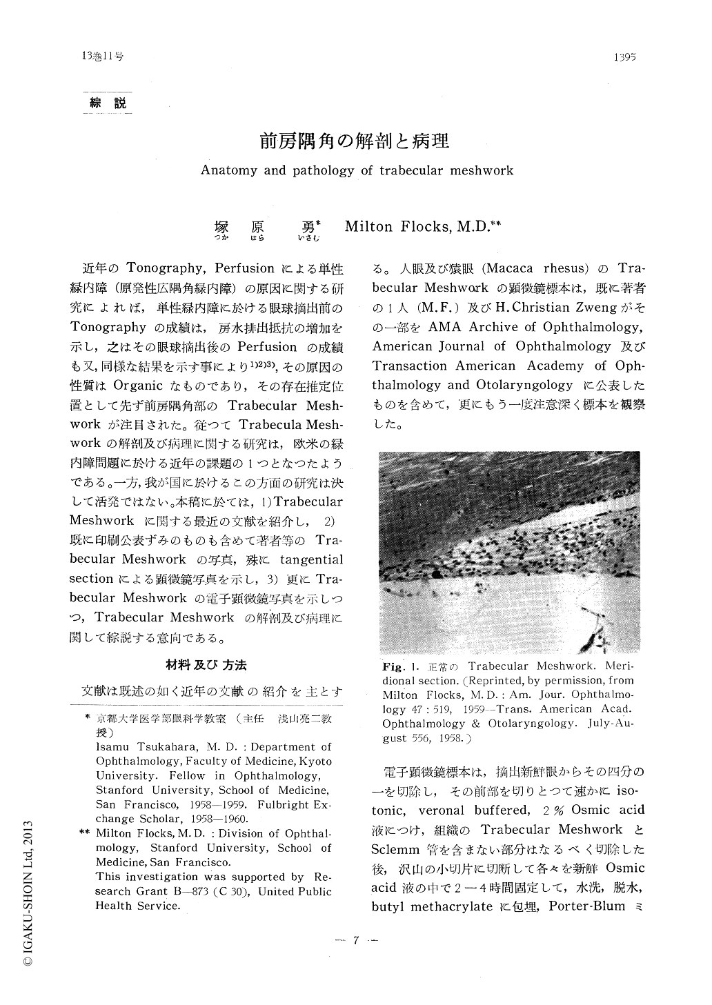
近年のTonography,Perfusionによる単性緑内障(原発性広隅角緑内障)の原因に関する研究によれば,単性緑内障に於ける眼球摘出前のTonographyの成績は,房水排出抵抗の増加を示し,之はその眼球摘出後のPerfusionの成績も又,同様な結果を示す事により1)2)3),その原因の性質はOrganicなものであり,その存在推定位置として先ず前房隅角部のTrabecular Mesh-workが注目された。従つてTrabecula Mesh-workの解剖及び病理に関する研究は,欧米の緑内障問題に於ける近年の課題の1つとなつたようである。一方,我が国に於けるこの方面の研究は決して活発ではない。本稿に於ては,1) TrabecularMeshworkに関する最近の文献を紹介し,2)既に印刷公表ずみのものも含めて著者等のTra-becular Meshworkの写真,殊にtangentialsectionによる顕微鏡写真を示し,3)更にTra-becular Meshworkの電子顕微鏡写真を示しつつ,Trabecular Meshworkの解剖及び病理に関して綜説する意向である。
This is an introduction of anatomy and pathology of the trabecular meshwork. The authors review recent literatures on this subject and describe anatomy of the meshwork in tangential section. They show pictures of the meshwork in tangential section and recent electron microscopic pictures of the meshwork. Most of the pictures of the trabecular meshwork in tangential section which are shown in this paper were published in AMA Archive of Ophthalmology, American Journal of Ophthalmology and Transaction American Academy of Ophthalmology and Otolaryngology by one of the authors (M.F.).

Copyright © 1959, Igaku-Shoin Ltd. All rights reserved.


