Japanese
English
- 有料閲覧
- Abstract 文献概要
- 1ページ目 Look Inside
- 参考文献 Reference
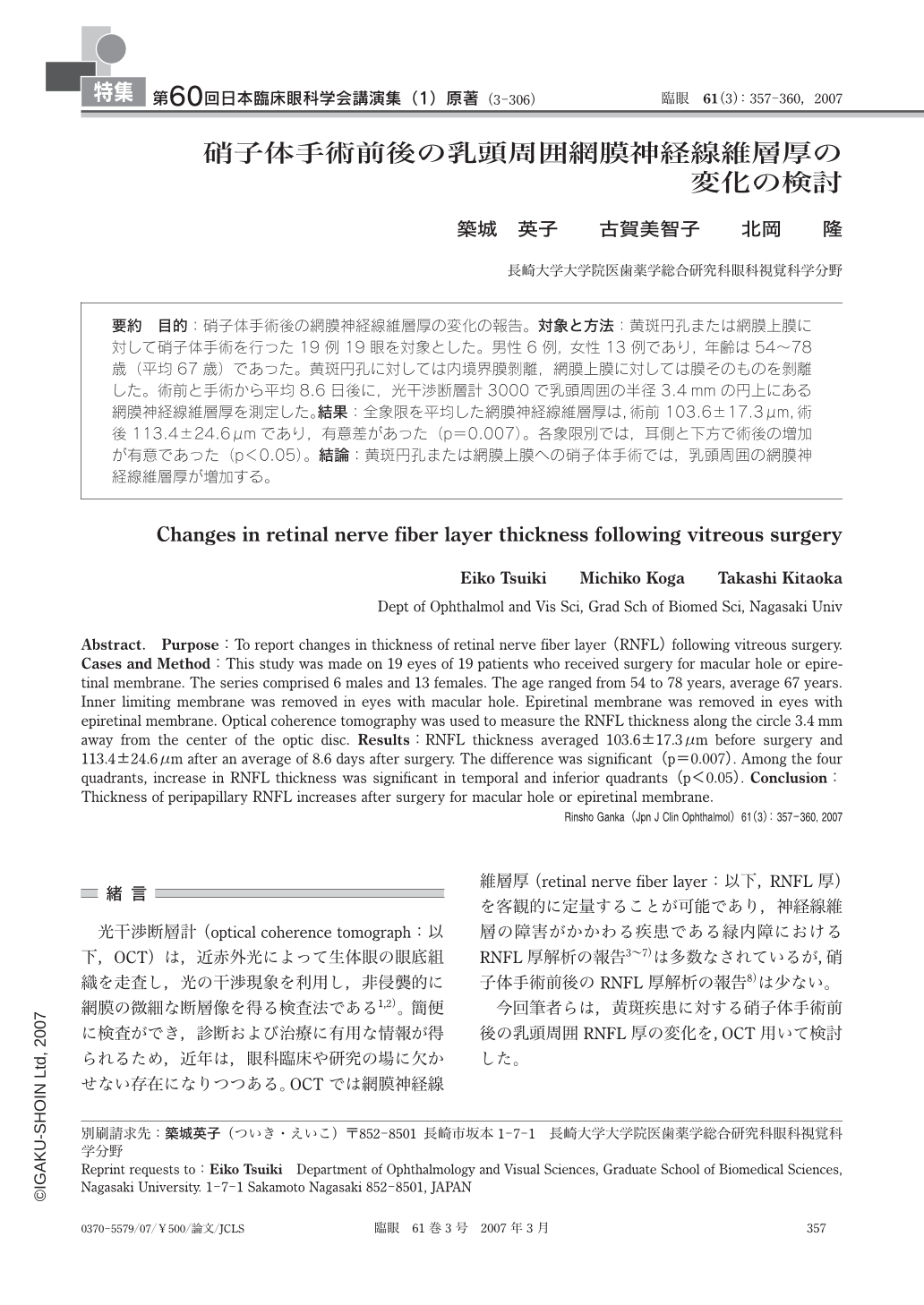
要約 目的:硝子体手術後の網膜神経線維層厚の変化の報告。対象と方法:黄斑円孔または網膜上膜に対して硝子体手術を行った19例19眼を対象とした。男性6例,女性13例であり,年齢は54~78歳(平均67歳)であった。黄斑円孔に対しては内境界膜剝離,網膜上膜に対しては膜そのものを剝離した。術前と手術から平均8.6日後に,光干渉断層計3000で乳頭周囲の半径3.4mmの円上にある網膜神経線維層厚を測定した。結果:全象限を平均した網膜神経線維層厚は,術前103.6±17.3μm,術後113.4±24.6μmであり,有意差があった(p=0.007)。各象限別では,耳側と下方で術後の増加が有意であった(p<0.05)。結論:黄斑円孔または網膜上膜への硝子体手術では,乳頭周囲の網膜神経線維層厚が増加する。
Abstract. Purpose:To report changes in thickness of retinal nerve fiber layer(RNFL)following vitreous surgery. Cases and Method:This study was made on 19 eyes of 19 patients who received surgery for macular hole or epiretinal membrane. The series comprised 6 males and 13 females. The age ranged from 54 to 78 years,average 67 years. Inner limiting membrane was removed in eyes with macular hole. Epiretinal membrane was removed in eyes with epiretinal membrane. Optical coherence tomography was used to measure the RNFL thickness along the circle 3.4mm away from the center of the optic disc. Results:RNFL thickness averaged 103.6±17.3μm before surgery and 113.4±24.6μm after an average of 8.6 days after surgery. The difference was significant(p=0.007). Among the four quadrants,increase in RNFL thickness was significant in temporal and inferior quadrants(p<0.05). Conclusion:Thickness of peripapillary RNFL increases after surgery for macular hole or epiretinal membrane.

Copyright © 2007, Igaku-Shoin Ltd. All rights reserved.


