Japanese
English
シンポジウム 骨盤臼蓋の発育
保存的治療後の臼蓋発育
Radiological Study of Growth and Development of the Acetabulum in Non Operatively Treated Congenital Dislocation Hip
池田 定倫
1
,
岩崎 勝郎
1
,
鈴木 良平
1
Sadamichi Ikeda
1
1長崎大学医学部整形外科学教室
1Department of Orthopaedic Surgery, Nagasaki University, School of Medicine
キーワード:
先天股脱の保存的治療
,
conservation treatment of congenital dislocative of the hip
,
臼蓋の発育
,
growth and development of the acetabulum
,
寛骨臼の形態と治療成績
,
last screening by acetabular shape
,
臼蓋の発育のX線学的経過
,
radiological assessment of growth of the acetabulum
Keyword:
先天股脱の保存的治療
,
conservation treatment of congenital dislocative of the hip
,
臼蓋の発育
,
growth and development of the acetabulum
,
寛骨臼の形態と治療成績
,
last screening by acetabular shape
,
臼蓋の発育のX線学的経過
,
radiological assessment of growth of the acetabulum
pp.51-57
発行日 1986年1月25日
Published Date 1986/1/25
DOI https://doi.org/10.11477/mf.1408907327
- 有料閲覧
- Abstract 文献概要
- 1ページ目 Look Inside
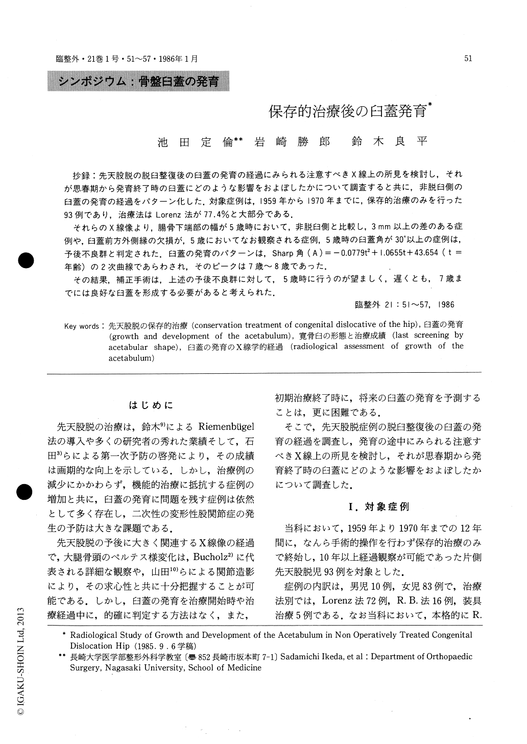
抄録:先天股脱の脱臼整復後の臼蓋の発育の経過にみられる注意すべきX線上の所見を検討し,それが思春期から発育終了時の臼蓋にどのような影響をおよぼしたかについて調査すると共に,非脱臼側の臼蓋の発育の経過をパターン化した.対象症例は,1959年から1970年までに,保存的治療のみを行った93例であり,治療法はLorenz法が77.4%と大部分である.
それらのX線像より,腸骨下端部の幅が5歳時において,非脱臼側と比較し,3mm以上の差のある症例や,臼蓋前方外側縁の欠損が,5歳においてなお観察される症例,5歳時の臼蓋角が30゜以上の症例は,予後不良群と判定された.臼蓋の発育のパターンは,Sharp角(A)=-0.0779t2+1.0655t+43.654(t=年齢)の2次曲線であらわされ,そのピークは7歳〜8歳であった.

Copyright © 1986, Igaku-Shoin Ltd. All rights reserved.


