Japanese
English
- 有料閲覧
- Abstract 文献概要
- 1ページ目 Look Inside
- 参考文献 Reference
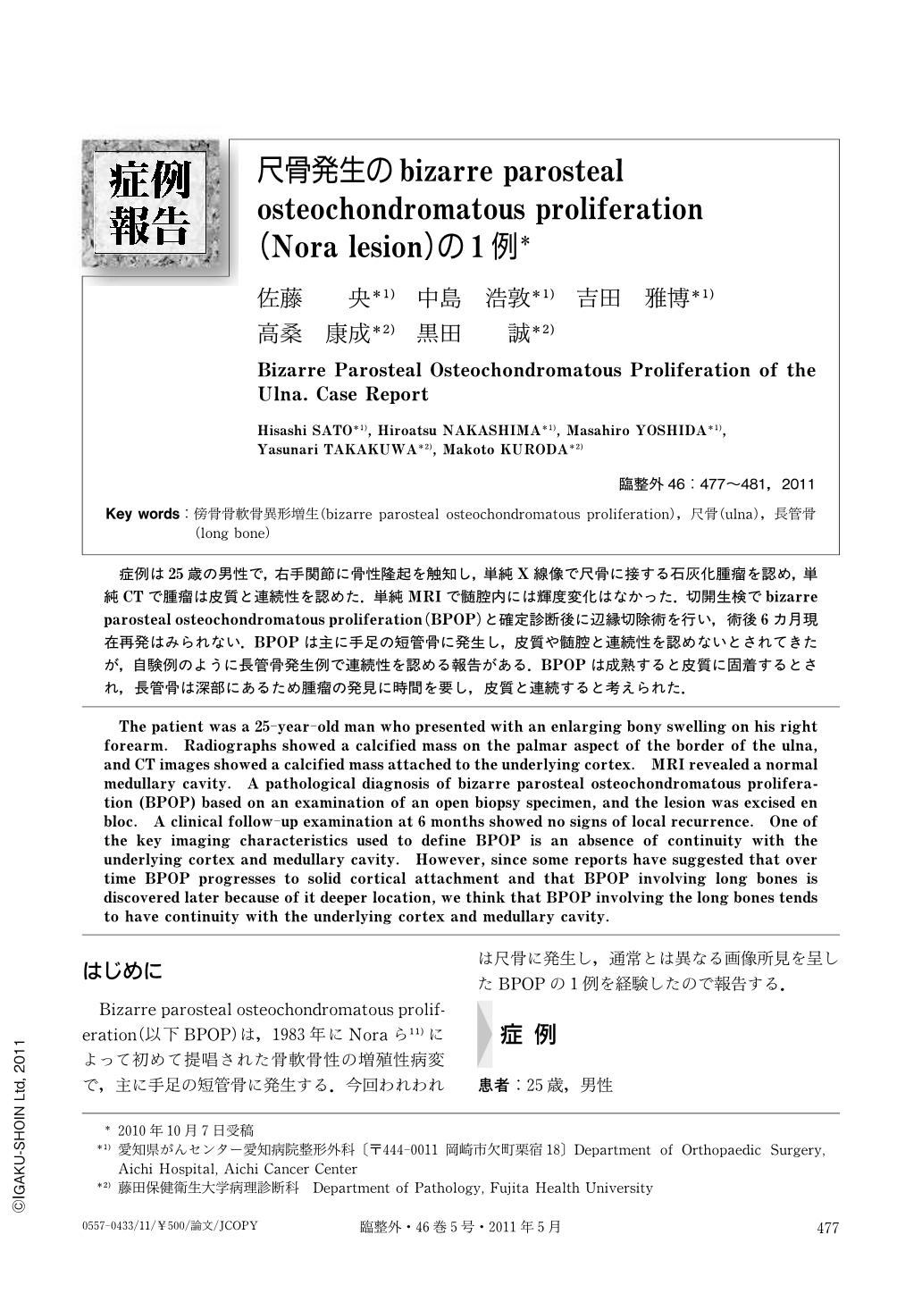
症例は25歳の男性で,右手関節に骨性隆起を触知し,単純X線像で尺骨に接する石灰化腫瘤を認め,単純CTで腫瘤は皮質と連続性を認めた.単純MRIで髄腔内には輝度変化はなかった.切開生検でbizarre parosteal osteochondromatous proliferation(BPOP)と確定診断後に辺縁切除術を行い,術後6カ月現在再発はみられない.BPOPは主に手足の短管骨に発生し,皮質や髄腔と連続性を認めないとされてきたが,自験例のように長管骨発生例で連続性を認める報告がある.BPOPは成熟すると皮質に固着するとされ,長管骨は深部にあるため腫瘤の発見に時間を要し,皮質と連続すると考えられた.
The patient was a 25-year-old man who presented with an enlarging bony swelling on his right forearm. Radiographs showed a calcified mass on the palmar aspect of the border of the ulna, and CT images showed a calcified mass attached to the underlying cortex. MRI revealed a normal medullary cavity. A pathological diagnosis of bizarre parosteal osteochondromatous proliferation (BPOP) based on an examination of an open biopsy specimen, and the lesion was excised en bloc. A clinical follow-up examination at 6 months showed no signs of local recurrence. One of the key imaging characteristics used to define BPOP is an absence of continuity with the underlying cortex and medullary cavity. However, since some reports have suggested that over time BPOP progresses to solid cortical attachment and that BPOP involving long bones is discovered later because of it deeper location, we think that BPOP involving the long bones tends to have continuity with the underlying cortex and medullary cavity.

Copyright © 2011, Igaku-Shoin Ltd. All rights reserved.


