Japanese
English
- 有料閲覧
- Abstract 文献概要
- 1ページ目 Look Inside
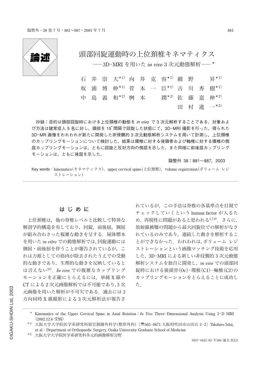
抄録:目的は頭部回旋時における上位頚椎の動態をin vivoで3次元解析することである.対象および方法は健常成人5名に対し,頭部を15°間隔で回旋した状態にて,3D-MRI撮影を行った.得られた3D-MR画像をわれわれが新たに開発した非侵襲的3次元動態解析システムを用いて計測し,上位頚椎のカップリングモーションについて検討した.結果は環椎に対する後頭骨および軸椎に対する環椎の側屈カップリングモーションは,ともに回旋と反対方向の側屈を示した.また同様に前後屈カップリングモーションは,ともに後屈を示した.
The coupling motion of the upper cervical spine has been investigated in vitro using cadavers, and in vivo using bi-planar radiographs ; neither method allows following of real motion. We developed a unique motion analysis system and successfully described the in vivo three-dimensional motion of the upper cervical spine using this system. Three-dimensional MR images of the upper cervical spine were taken for 5 healthy volunteers, using a 1.0-T imager in progressive 15° steps of head rotation. The segmented three-dimensional MR image of each vertebra in the neutral position was superimposed over images taken at other positions, using voxel-based registration. Relative motion of the upper cervical spine was measured and described with 6 freedoms, using rigid body Euler angles and translations. At maximum head rotation, average axial rotation angle was 2.4° between Oc and C1, and 35.9° between C1 and C2. Coupled lateral bending with axial rotation was observed in the direction opposite to that of axial rotation at Oc-C1 and C1-C2. Coupled extension with axial rotation occurred at Oc-C1 and C1-C2. The present results are in good agreement with those of an in vitro study by Panjabi.

Copyright © 2003, Igaku-Shoin Ltd. All rights reserved.


