Japanese
English
- 有料閲覧
- Abstract 文献概要
- 1ページ目 Look Inside
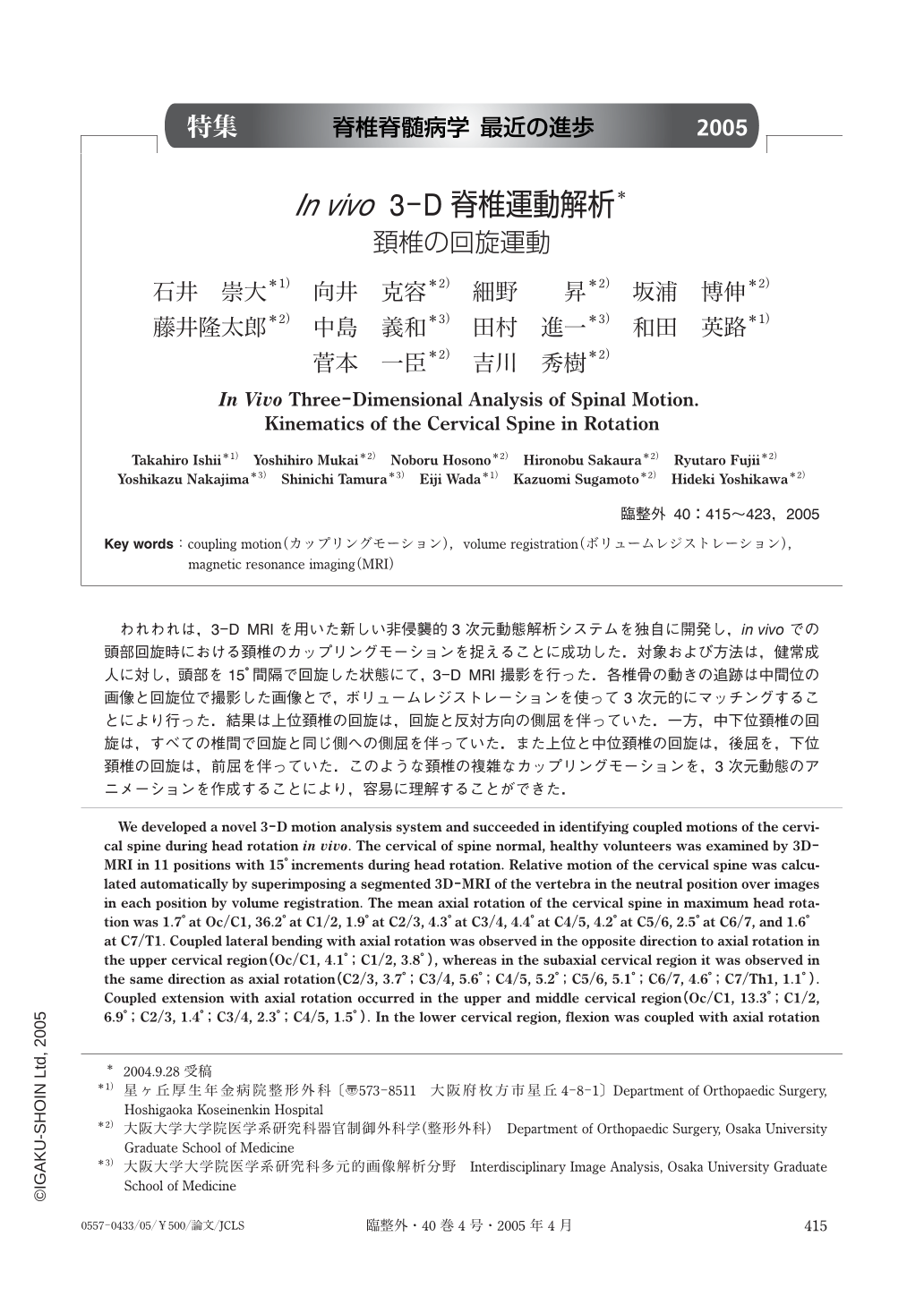
われわれは,3-D MRIを用いた新しい非侵襲的3次元動態解析システムを独自に開発し,in vivoでの頭部回旋時における頚椎のカップリングモーションを捉えることに成功した.対象および方法は,健常成人に対し,頭部を15°間隔で回旋した状態にて,3-D MRI撮影を行った.各椎骨の動きの追跡は中間位の画像と回旋位で撮影した画像とで,ボリュームレジストレーションを使って3次元的にマッチングすることにより行った.結果は上位頚椎の回旋は,回旋と反対方向の側屈を伴っていた.一方,中下位頚椎の回旋は,すべての椎間で回旋と同じ側への側屈を伴っていた.また上位と中位頚椎の回旋は,後屈を,下位頚椎の回旋は,前屈を伴っていた.このような頚椎の複雑なカップリングモーションを,3次元動態のアニメーションを作成することにより,容易に理解することができた.
We developed a novel 3-D motion analysis system and succeeded in identifying coupled motions of the cervical spine during head rotation in vivo. The cervical of spine normal, healthy volunteers was examined by 3D-MRI in 11 positions with 15° increments during head rotation. Relative motion of the cervical spine was calculated automatically by superimposing a segmented 3D-MRI of the vertebra in the neutral position over images in each position by volume registration. The mean axial rotation of the cervical spine in maximum head rotation was 1.7° at Oc/C1, 36.2° at C1/2, 1.9° at C2/3, 4.3° at C3/4, 4.4° at C4/5, 4.2° at C5/6, 2.5° at C6/7, and 1.6° at C7/T1. Coupled lateral bending with axial rotation was observed in the opposite direction to axial rotation in the upper cervical region (Oc/C1, 4.1°;C1/2, 3.8°), whereas in the subaxial cervical region it was observed in the same direction as axial rotation (C2/3, 3.7°;C3/4, 5.6°;C4/5, 5.2°;C5/6, 5.1°;C6/7, 4.6°;C7/Th1, 1.1°). Coupled extension with axial rotation occurred in the upper and middle cervical region (Oc/C1, 13.3°;C1/2, 6.9°;C2/3, 1.4°;C3/4, 2.3°;C4/5, 1.5°). In the lower cervical region, flexion was coupled with axial rotation (C5/6, 0.9°;C6/7, 2.4°;C7/T1, 3.0°). Our system not only facilitates mathematical descriptions of 3D motion, but 3D visualization of the information, and made it possible to easily understand complicated coupled motions of the cervical spine.

Copyright © 2005, Igaku-Shoin Ltd. All rights reserved.


