Japanese
English
- 有料閲覧
- Abstract 文献概要
- 1ページ目 Look Inside
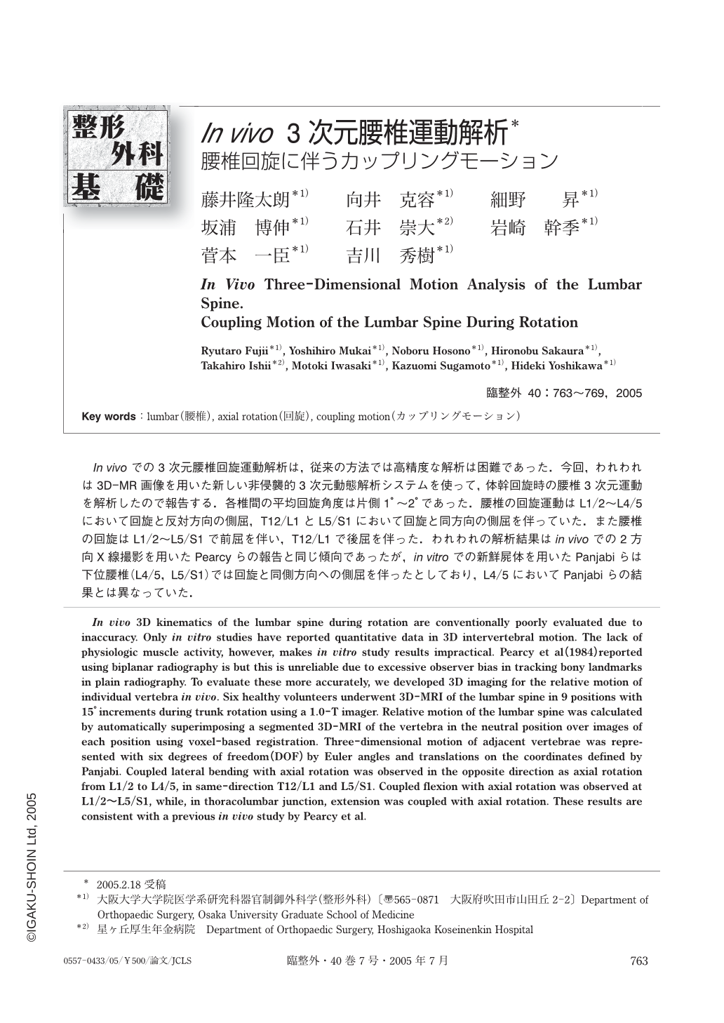
In vivoでの3次元腰椎回旋運動解析は,従来の方法では高精度な解析は困難であった.今回,われわれは3D-MR画像を用いた新しい非侵襲的3次元動態解析システムを使って,体幹回旋時の腰椎3次元運動を解析したので報告する.各椎間の平均回旋角度は片側1°~2°であった.腰椎の回旋運動はL1/2~L4/5において回旋と反対方向の側屈,T12/L1とL5/S1において回旋と同方向の側屈を伴っていた.また腰椎の回旋はL1/2~L5/S1で前屈を伴い,T12/L1で後屈を伴った.われわれの解析結果はin vivoでの2方向X線撮影を用いたPearcyらの報告と同じ傾向であったが,in vitroでの新鮮屍体を用いたPanjabiらは下位腰椎(L4/5,L5/S1)では回旋と同側方向への側屈を伴ったとしており,L4/5においてPanjabiらの結果とは異なっていた.
In vivo 3D kinematics of the lumbar spine during rotation are conventionally poorly evaluated due to inaccuracy. Only in vitro studies have reported quantitative data in 3D intervertebral motion. The lack of physiologic muscle activity, however, makes in vitro study results impractical. Pearcy et al (1984) reported using biplanar radiography is but this is unreliable due to excessive observer bias in tracking bony landmarks in plain radiography. To evaluate these more accurately, we developed 3D imaging for the relative motion of individual vertebra in vivo. Six healthy volunteers underwent 3D-MRI of the lumbar spine in 9 positions with 15° increments during trunk rotation using a 1.0-T imager. Relative motion of the lumbar spine was calculated by automatically superimposing a segmented 3D-MRI of the vertebra in the neutral position over images of each position using voxel-based registration. Three-dimensional motion of adjacent vertebrae was represented with six degrees of freedom (DOF) by Euler angles and translations on the coordinates defined by Panjabi. Coupled lateral bending with axial rotation was observed in the opposite direction as axial rotation from L1/2 to L4/5, in same-direction T12/L1 and L5/S1. Coupled flexion with axial rotation was observed at L1/2~L5/S1, while, in thoracolumbar junction, extension was coupled with axial rotation. These results are consistent with a previous in vivo study by Pearcy et al.

Copyright © 2005, Igaku-Shoin Ltd. All rights reserved.


