Japanese
English
- 有料閲覧
- Abstract 文献概要
- 1ページ目 Look Inside
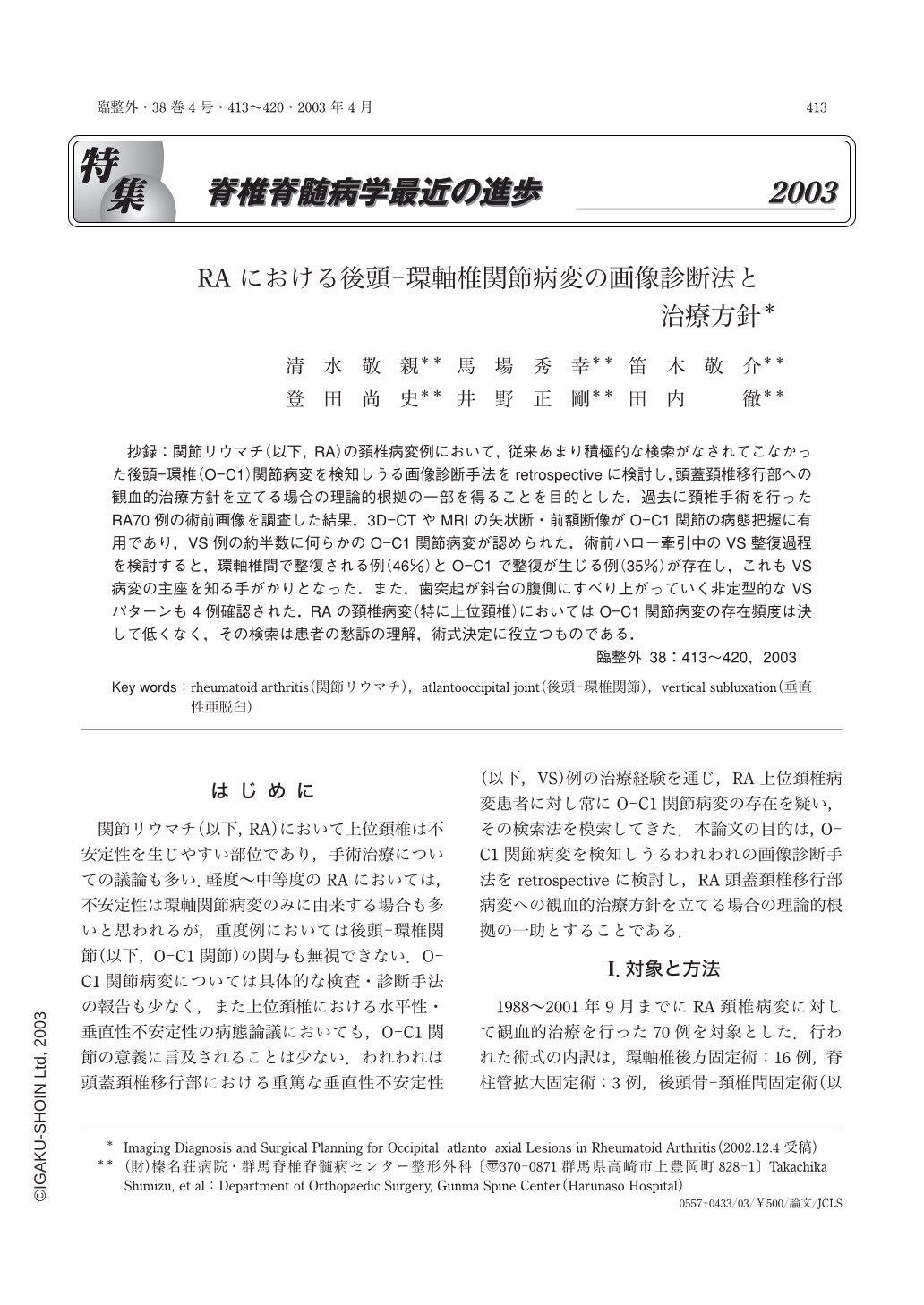
抄録:関節リウマチ(以下,RA)の頚椎病変例において,従来あまり積極的な検索がなされてこなかった後頭-環椎(O-C1)関節病変を検知しうる画像診断手法をretrospectiveに検討し,頭蓋頚椎移行部への観血的治療方針を立てる場合の理論的根拠の一部を得ることを目的とした.過去に頚椎手術を行ったRA70例の術前画像を調査した結果,3D-CTやMRIの矢状断・前額断像がO-C1関節の病態把握に有用であり,VS例の約半数に何らかのO-C1関節病変が認められた.術前ハロー牽引中のVS整復過程を検討すると,環軸椎間で整復される例(46%)とO-C1で整復が生じる例(35%)が存在し,これもVS病変の主座を知る手がかりとなった.また,歯突起が斜台の腹側にすべり上がっていく非定型的なVSパターンも4例確認された.RAの頚椎病変(特に上位頚椎)においてはO-C1関節病変の存在頻度は決して低くなく,その検索は患者の愁訴の理解,術式決定に役立つものである.
The purpose of this study was to identify atlanto-occipital (O-C1) joint pathology in rheumatoid arthritis of the cervical spine and to utilize the information obtained for surgical planning. Several neuroradiological findings in seventy cervical spine surgery cases were retrospectively reviewed. Multidirectional MRI sections and 3D-CT are useful tools for identifying craniocervical pathology, and they revealed atlanto-occipital abnormalities in over 60% of the cases in this series. In the vertical subluxation (VS) cases, vertical realignment (reduction) was achieved at O-C1 in 35% of the cases and at C1-C2 in 46% of the cases, thereby providing clues to the main levels of pathology in VS cases. Atypical VS patterns were also found in 4 cases. O-C1 lesions should be accurately identified in every patient with a rheumatoid disorder of the cervical spine, and surgical options should be considered based on the pathology of O-C1 lesions.

Copyright © 2003, Igaku-Shoin Ltd. All rights reserved.


