Japanese
English
- 有料閲覧
- Abstract 文献概要
- 1ページ目 Look Inside
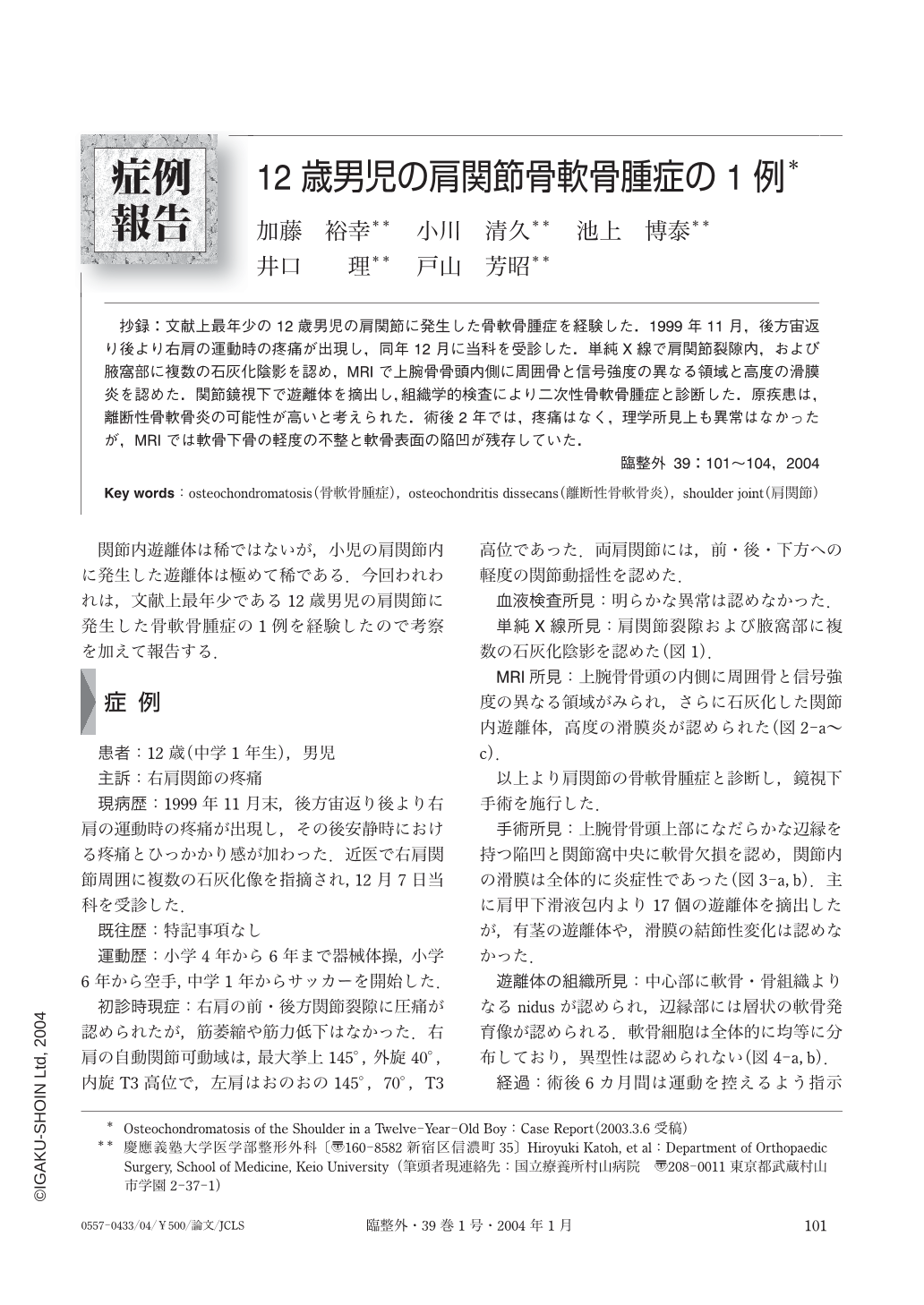
抄録:文献上最年少の12歳男児の肩関節に発生した骨軟骨腫症を経験した.1999年11月,後方宙返り後より右肩の運動時の疼痛が出現し,同年12月に当科を受診した.単純X線で肩関節裂隙内,および腋窩部に複数の石灰化陰影を認め,MRIで上腕骨骨頭内側に周囲骨と信号強度の異なる領域と高度の滑膜炎を認めた.関節鏡視下で遊離体を摘出し,組織学的検査により二次性骨軟骨腫症と診断した.原疾患は,離断性骨軟骨炎の可能性が高いと考えられた.術後2年では,疼痛はなく,理学所見上も異常はなかったが,MRIでは軟骨下骨の軽度の不整と軟骨表面の陥凹が残存していた.
We present a rare case of secondary osteochondromatosis in the shoulder joint of a twelve-year-old boy, and discuss the possible etiology of the condition. In December 1999, a twelve-year-old boy presented with pain in the right shoulder after conducting a back flip a month earlier. Radiographs revealed multiple calcified bodies in the shoulder joint and the axillary area. MRI revealed severe synovitis and an edematous lesion in the medial humeral head. An arthroscopic extraction of the calcified bodies was performed and histological examination of the extracted bodies led to the diagnosis of secondary osteochondromatosis. Of the numerous conditions that may lead to secondary osteochondromatosis, osteochondritis dissecans may have been the precipitating condition in this case. Two years after surgery, the patient has no complaints of pain, and there are no remarkable findings upon physical examination. MRI revealed that an irregularity in the subchondral bone and a chondral depression remained in the medial humeral head. There have been no recurrences of free bodies during the two year follow-up period.

Copyright © 2004, Igaku-Shoin Ltd. All rights reserved.


