Japanese
English
- 有料閲覧
- Abstract 文献概要
- 1ページ目 Look Inside
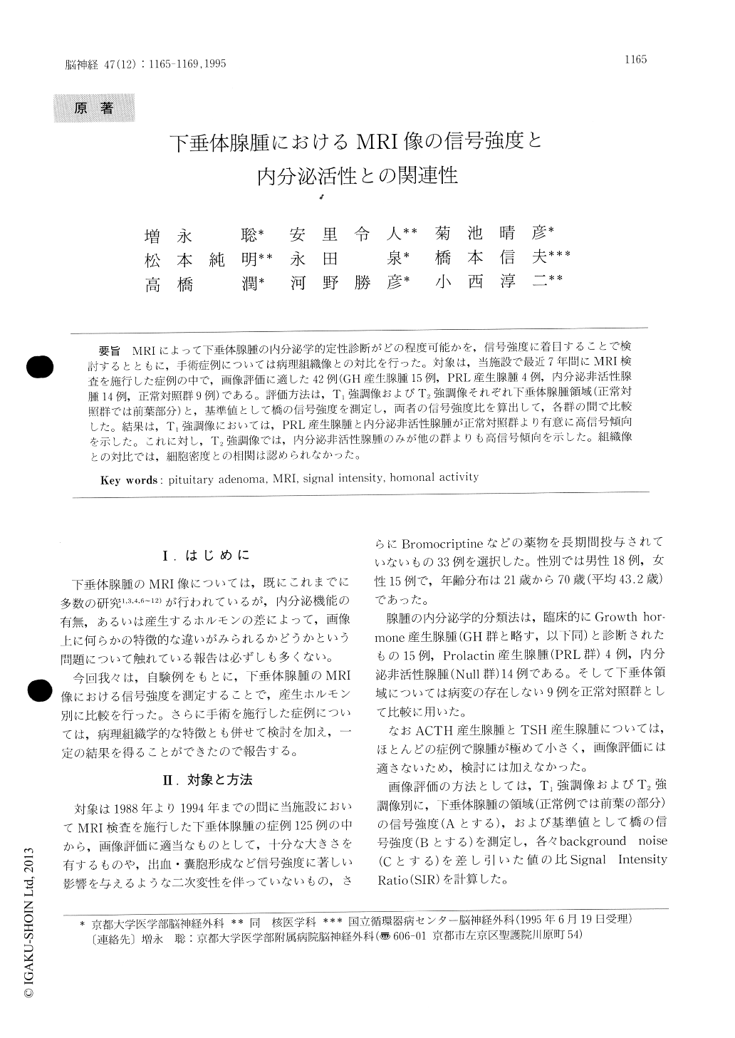
MRIによって下垂体腺腫の内分泌学的定性診断がどの程度可能かを,信号強度に着目することで検討するとともに,手術症例については病理組織像との対比を行った。対象は,当施設で最近7年間にMRI検査を施行した症例の中で,画像評価に適した42例(GH産生腺腫15例,PRL産生腺腫4例,内分泌非活性腺腫14例,正常対照群9例)である。評価方法は,T1強調像およびT2強調像それぞれ下垂体腺腫領域(正常対照群では前葉部分)と,基準値として橋の信号強度を測定し,両者の信号強度比を算出して,各群の間で比較した。結果は,T1強調像においては,PRL産生腺腫と内分泌非活性腺腫が正常対照群より有意に高信号傾向を示した。これに対し,T2強調像では,内分泌非活性腺腫のみが他の群よりも高信号傾向を示した。組織像との対比では,細胞密度との相関は認められなかった。
Many studies in Magnetic Resonance Imaging (MRI) of pituitary adenomas are already perform-ed. However, few reports exist about MRI findings of pituitary adenomas with reference to the hor-monal activity, therefore, we evaluated this problem on the viewpoint of the signal intensity in MRI and pathological features. Fifteen patients with growth hormone producing adenoma (GH-group), 6 patients with prolactin producing adenoma (PRL-group), 15 patients with endocrinologically non-functioning adenoma (Null-group) and 9 cases with normal pituitary gland (normal control group) were examined.

Copyright © 1995, Igaku-Shoin Ltd. All rights reserved.


