Japanese
English
- 有料閲覧
- Abstract 文献概要
- 1ページ目 Look Inside
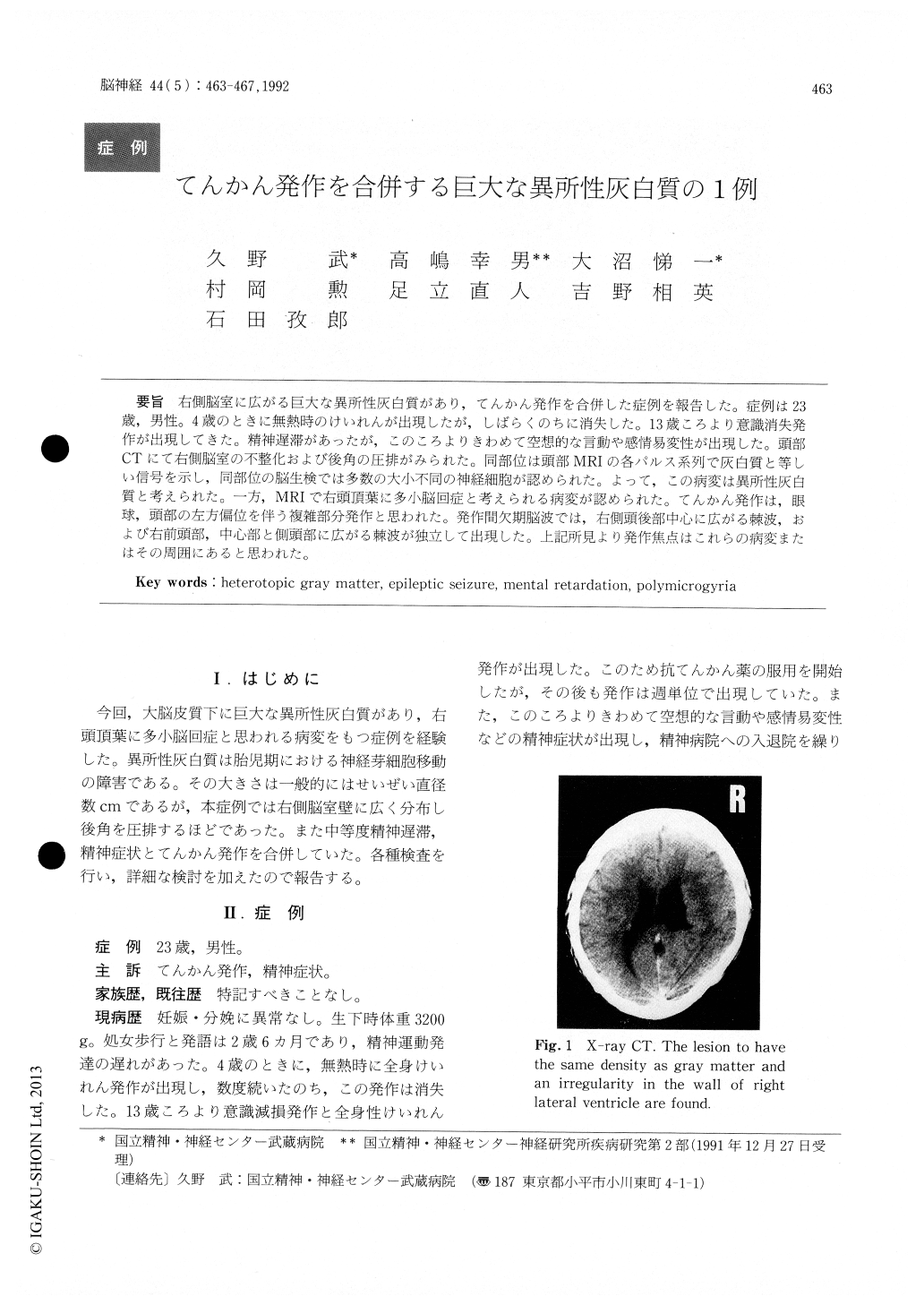
右側脳室に広がる巨大な異所性灰白質があり,てんかん発作を合併した症例を報告した。症例は23歳,男性。4歳のときに無熱時のけいれんが出現したが,しばらくのちに消失した。13歳ころより意識消失発作が出現してきた。精神遅滞があったが,このころよりきわめて空想的な言動や感情易変性が出現した。頭部CTにて右側脳室の不整化および後角の圧排がみられた。同部位は頭部MRIの各パルス系列で灰白質と等しい信号を示し,同部位の脳生検では多数の大小不同の神経細胞が認められた。よって,この病変は異所性灰白質と考えられた。一方,MRIで右頭頂葉に多小脳回症と考えられる病変が認められた。てんかん発作は,眼球,頭部の左方偏位を伴う複雑部分発作と思われた。発作間欠期脳波では,右側頭後部中心に広がる棘波,および右前頭部,中心部と側頭部に広がる棘波が独立して出現した。上記所見より発作焦点はこれらの病変またはその周囲にあると思われた。
We report a patient with partial seizure and gigantic heterotopic gray matter. A 23-year-old young man was admitted to our hospital with corn-plaints of frequent epileptic seizures and psychiatric symptoms. There was psychomotor delay in infancy. At the age of 4 years, afebrile convulsions appeared on several occasions. Seizures character-ized by a lapse of consciousness started at the age of 13 years. He often stayed in a fantasy world and became very emotional at such time. Cranial CT demostrated an irregularity in the wall of right lateral ventricle and disappearance of the posterior horn on the same side. This lesion, adjacent to that wall, had a signal intensity that was similar to that of the gray matter on each sequence in MRI. His-topathology of this lesion showed a number of large and small neurons. Therefore, heterotopic gray matter was diagnosed. MRI demonstrated wide cortices suggesting polymicrogyria in the right par-ietal lobe. Complex partial seizures with eye devia-tion to the left were recognized. Interictal EEG showed frequent high voltage spikes in the right temporal, fronto-temporal and parieto-occipital areas independently. Therefore, epileptic foci were thought to exist in or around those lesions.

Copyright © 1992, Igaku-Shoin Ltd. All rights reserved.


