Japanese
English
- 有料閲覧
- Abstract 文献概要
- 1ページ目 Look Inside
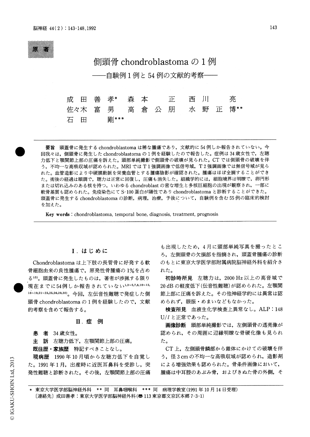
頭蓋骨に発生するchondroblastomaは稀な腫瘍であり,文献的に54例しか報告されていない。今回我々は,側頭骨に発生したchondroblastomaの1例を経験したので報告した。症例は34歳女性で,左聴力低下と顎関節上部の圧痛を訴えた。頭部単純撮影で側頭骨の破壊が見られた。CTでは側頭骨の破壊を伴う,不均一な高吸収域が認められた。MRIではT1強調画像で低信号域,T2強調画像では無信号域が見られた。血管造影により中硬膜動脈を栄養血管とする腫瘍陰影が確認された。腫瘍はほぼ全摘することができた。術後の経過は順調で,聴力は正常に回復し,圧痛も消失した。組織学的には,細胞境界は明瞭で,卵円形または切れ込みのある核を持つ,いわゆるchondroblastの密な増生と多核巨細胞の出現が観察され,一部に軟骨基質も認められた。免疫染色にてS−100蛋白が陽性でありchondroblastomaと診断することができた。頭蓋骨に発生するchondroblastomaの診断,病理,治療,予後について,自験例を含む55例の臨床的検討を加えた。
Chondroblastoma of the skull is a rare tumor and only 54 cases have been reported to date. A case of chondroblastoma arising from the squamous part of the left tempomal bone is reported. A 34-year-oldwoman had 6-month history of left conductive hear-ing disturbance and tenderness in the left temporal region. Plain skull X-ray showed a well demarcated osteolytic lesion in the temporal bone. CT demon-strated a heterogeneously high density mass, with enhancement. T 1-weighted MRI showed a low intensity mass while T2-weighted images showed no signal area. The left external carotid angiograms showed a marked staining supplied by the left middle meningial artery. This tumor grew destroy-ing the left temporal squama and pyramidal bone, and extended to the external auditory canal and the middle ear cavity. The tumor was subtotally resect-ed. Histologically, this tumor consisted of clusters of round or polygonal chondroblasts with oval or grooved nuclei and well-defined cell border. Multinucleated giant cells were also observed. Chondroid matrix was found in some areas. Im-munohistochemically, the tumor cells were positve for S-100 protein. These findings lead us to the diagnosis of chondroblastoma. The diagnosis, his-tology, therapy, and prognosis of chondroblastoma are discussed including the review of 54 cases in the literature.

Copyright © 1992, Igaku-Shoin Ltd. All rights reserved.


