Japanese
English
- 有料閲覧
- Abstract 文献概要
- 1ページ目 Look Inside
- 参考文献 Reference
Ⅰ.はじめに
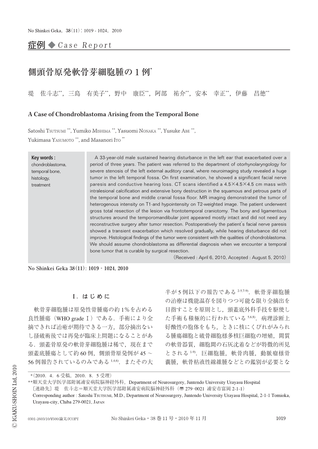
軟骨芽細胞腫は原発性骨腫瘍の約1%を占める良性腫瘍(WHO grade Ⅰ)である.手術により全摘できれば治癒が期待できる一方,部分摘出ないし掻破術後では再発が臨床上問題になることがある.頭蓋骨原発の軟骨芽細胞腫は稀で,現在まで頭蓋底腫瘍として約60例,側頭骨原発例が45~56例報告されているのみである1,4,6).またその大半が5例以下の報告である2-5,7-9).軟骨芽細胞腫の治療は機能温存を図りつつ可能な限り全摘出を目指すことを原則とし,頭蓋底外科手技を駆使した手術も積極的に行われている5,6,8).病理診断上好酸性の胞体をもち,ときに核にくびれがみられる腫瘍細胞と破骨細胞様多核巨細胞の増殖,間質の軟骨器質,細胞間の石灰沈着などが特徴的所見とされる1-9).巨細胞腫,軟骨肉腫,動脈瘤様骨囊腫,軟骨粘液性線維腫などとの鑑別が必要となるが,近年S-100染色の診断上の有用性が認識されるようになった1,9).今回われわれは伝音性聴力障害で発症,手術により治療した中頭蓋窩軟骨芽細胞腫の症例を経験したので報告する.
A 33-year-old male sustained hearing disturbance in the left ear that exacerbated over a period of three years. The patient was referred to the department of otorhynolaryngology for severe stenosis of the left external auditory canal, where neuroimaging study revealed a huge tumor in the left temporal fossa. On first examination, he showed a significant facial nerve paresis and conductive hearing loss. CT scans identified a 4.5×4.5×4.5 cm mass with intralesional calcification and extensive bony destruction in the squamous and petrous parts of the temporal bone and middle cranial fossa floor. MR imaging demonstrated the tumor of heterogenous intensity on T1-and hypointensity on T2-weighted image. The patient underwent gross total resection of the lesion via frontotemporal craniotomy. The bony and ligamentous structures around the temporomandibular joint appeared mostly intact and did not need any reconstructive surgery after tumor resection. Postoperatively the patient's facial nerve paresis showed a transient exacerbation which resolved gradually, while hearing disturbance did not improve. Histological findings of the tumor were consistent with the qualities of chondroblastoma. We should assume chondroblastoma as differential diagnosis when we encounter a temporal bone tumor that is curable by surgical resection.

Copyright © 2010, Igaku-Shoin Ltd. All rights reserved.


