Japanese
English
- 有料閲覧
- Abstract 文献概要
- 1ページ目 Look Inside
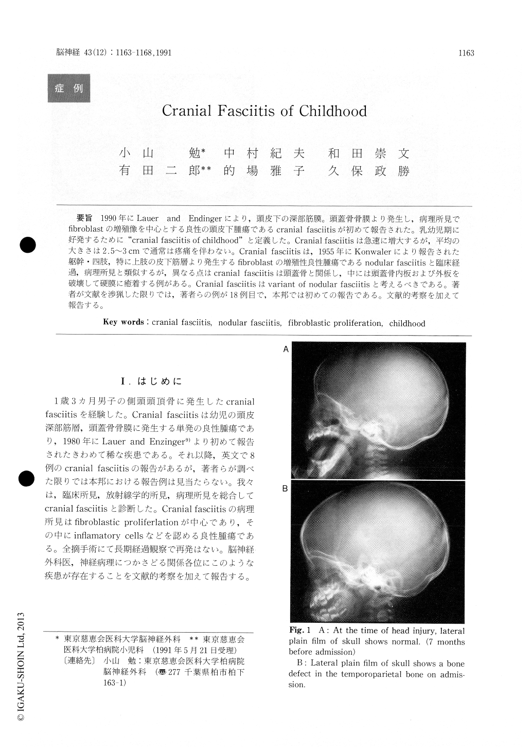
1990年にLauer and Endingerにより,頭皮下の深部筋膜。頭蓋骨骨膜より発生し,病理所見でfibroblastの増殖像を中心とする良性の頭皮下腫瘍であるcranial fasciitisが初めて報告された。乳幼児期に好発するために"cranial fasciitis of childhood"と定義した。Cranial fasciitisは急速に増大するが,平均の大きさは2.5〜3cmで通常は疼痛を伴わない。Cranial fasciitisは,1955年にKonwalerにより報告された躯幹・四肢,特に上肢の皮下筋層より発生するfibroblastの増殖性良性腫瘍であるnodular fasciitisと臨床経過,病理所見と類似するが,異なる点はcranial fasciitisは頭蓋骨と関係し,中には頭蓋骨内板および外板を破壊して硬膜に癒着する例がある。Cranial fasciitisはvariant of nodular fasciitisと考えるべきである。著者が文献を渉猟した限りでは,著者らの例が18例目で,本邦では初めての報告である。文献的考察を加えて報告する。
A rare case of cranial fasciitis in a 1-year-old boy arising in the temporoparietal bone has been de-scribed. In 1990, Lauer and Endinger first reported cranial fasciitis, which is a benign subcutaneous tumor of the head developig from the deep fascia or the cranial periosteum and showing a pathological finding characterized by proliferation of fibroblasts. They described this tumor as "cranial fasciitis of childhood" in view of a high incident in infants and child. Cranial fasciitis grows rapidly in the scalp without pain, but its mean size is 2.5-3cm. Cranial fasciitis is closely related a clinical course and pathological findings to nodular fasciitis, which is also a benign proliferative fibroblast tumor develop-ing from the subcutaneous muscular layers of the trunk and extremities (especially, the forearms), which was reported by Konwaler in 1955. However, cranial fasciitis differs from nodular fasciitis in that it is associated with the skull bone and, in many cases, the tumor destroys the inner and outer table of the skull and adheres to the dura mater. Cranialfasciitis should be considered to be a veriant of nodular fasciitis.

Copyright © 1991, Igaku-Shoin Ltd. All rights reserved.


