Japanese
English
- 有料閲覧
- Abstract 文献概要
- 1ページ目 Look Inside
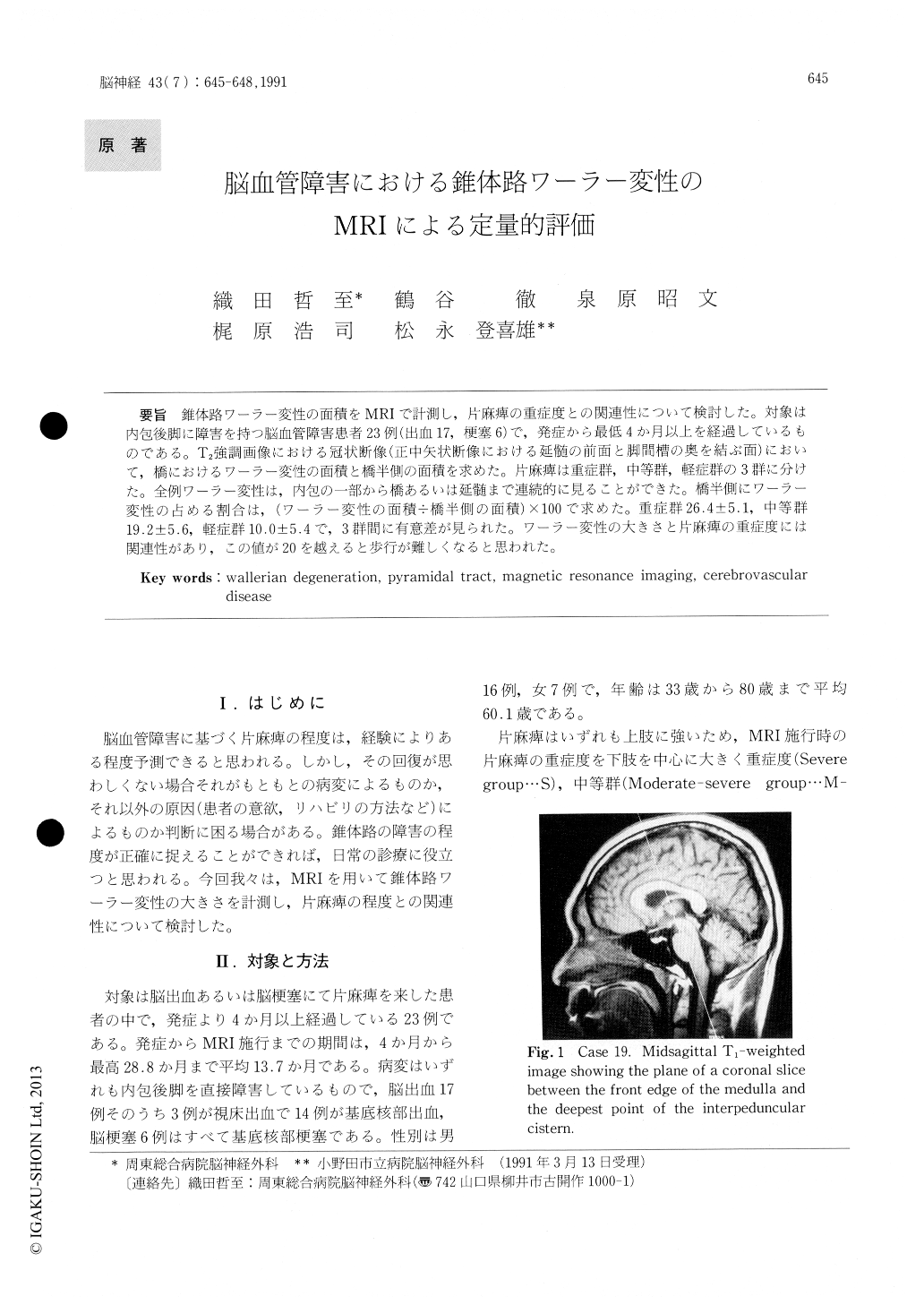
錐体路ワーラー変性の面積をMRIで計測し,片麻痺の重症度との関連性について検討した。対象は内包後脚に障害を持つ脳血管障害患者23例(出血17,梗塞6)で,発症から最低4か月以上を経過しているものである。T2強調画像における冠状断像(正中矢状断像における延髄の前面と脚間槽の奥を結ぶ面)において,橋におけるワーラー変性の面積と橋半側の面積を求めた。片麻痺は重症群,中等群,軽症群の3群に分けた。全例ワーラー変性は,内包の一部から橋あるいは延髄まで連続的に見ることができた。橋半側にワーラー変性の占める割合は,(ワーラー変性の面積÷橋半側の面積)×100で求めた。重症群26.4±5.1,中等群19.2±5.6,軽症群10.0±5.4で,3群間に有意差が見られた。ワーラー変性の大きさと片麻痺の重症度には関連性があり,この値が20を越えると歩行が難しくなると思われた。
Using magnetic resonance imaging, we studied 23 patients with motor deficit associated with cere-brovascular disease of the internal capsule. Accord-ing to the severity of the motor deficits, 23 patients were divided into three groups (severe group…9, moderately severe group…8, mild group…6). A coronal T2-weighted image was obtained along a straight line between the front edge of the medulla and the deepest point of the interpeduncular cistern in a midsagittal T1-weighted image. It was revealed that wallerian degeneration extended con-tinuously from part of the internal capsule down to the pons or medulla or the decussation in all patients. The area of wallerian degeneration in the pons and the area of half the pons were calculated from the coronal T2-weighted image. Moreover, the wallerian index was calculated as : (area of wallerian degeneration in pons÷area of half the pons)×100. Values of the wallerian index±SD were 26.4±5.1 in the severe group, 19.2±5.6 in the moderately severe group, and 10.0±5.4 in the mild group. There were significant differences among the three groups. We concluded that the area of wallerian degeneration was related to the severity of motor deficits.

Copyright © 1991, Igaku-Shoin Ltd. All rights reserved.


