Japanese
English
- 有料閲覧
- Abstract 文献概要
- 1ページ目 Look Inside
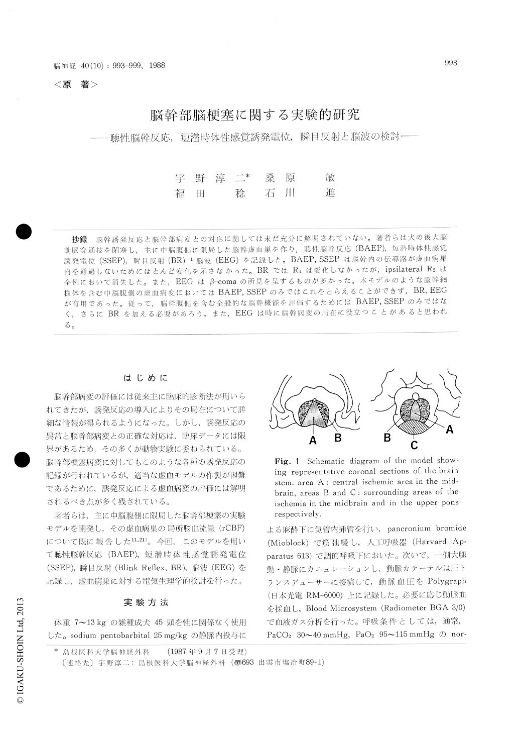
抄録 脳幹誘発反応と脳幹部病変との対応に関しては未だ充分に解明されていない。著者らは犬の後大脳動脈穿通枝を閉塞し,主に中脳腹側に限局した脳幹虚血巣を作り,聴性脳幹反応(BAEP),短潜時体性感覚誘発電位(SSEP),瞬目反射(BR)と脳波(EEG)を記録した。BAEP,SSEPは脳幹内の伝導路が虚血病巣内を通過しないためにほとんど変化を示さなかった。BRではR1は変化しなかったが,ipsilateral R2は全例において消失した。また,EEGはβ-comaの所見を呈するものが多かった。本モデルのような脳幹網様体を含む中脳腹側の虚血病変においてはBAEP,SSEPのみではこれをとらえることができず,BR,EEGが有用であった。従って,脳幹腹側を含む全般的な脳幹機能を評価するためにはBAEP,SSEPのみではなく,さらにBRを加える必要があろう。また,EEGは時に脳幹病変の局在に役立つことがあると思われる。
Assessment of the lesion in the brain stem by evoked potentials has not been well established. We have already developed a model of brain stem ischemia by occluding the perforators of the pos-terior cerebral arteries of the dog. The ischemic lesion locates mainly in the ventral side of the midbrain. Using this model, we assessed brain stem function by brain stem auditory evoked po-tential (BAEP), surface-and depth-recorded (in medial lemniscus) short latency somatosensory evoked potential (SSEP), blink reflex (BR) and electroencephalography (EEG), and investigated the correlation between the electrophysiological abnormalities and the lesion in the brain stem. The studies were performed for 6 hours after per-forator occlusion. Furthermore, depth-recorded SSEP and regional cerebral blood flow (rCBF) were measured under induced hypotension by with-drawal of arterial blood.
BAEP did not change in 13 of 16 animals. Surface-recorded SSEP remained unchanged in all 6 animals. The results are probably due to the fact that the lesion does not involve the auditory and somatosensory pathways and the accompany-ing events such as edema does not affect the both pathways. Depth-recorded SSEP remained unchan-ged after occlusion and did not disappear even when rCBF fell below 10 ml/100 g/min. It may be suggested that the threshold for electrical failure in the brain stem is much lower than that in the cortex. In BR, RI did not change but ipsilateral R2 became nearly invisible immediately after per-forator occlusion in all animals. The fact that the ischemic lesion did not involve the pons and dis-turbed reticular formation in the midbrain may probably account for the remaining of R1 and the disappearance of ipsilateral R2. In EEG, low voltage fast activity appeared in most animals despite semicomatose state after occlusion. This phenomenon is called "β-coma" in clinical cases.
We conclude that by evaluating brain stem func-tion with only BAEP and SSEP the lesion in the ventral side of the brain stem may be frequently overlooked and BR as well as BAEP and SSEP is essential in the assessment of the whole brain stem function. In addition, R2 in BR is suggested to be the indicator of the function in the reticular formation. Although the lesion responsible for the appearance of β-coma has not been well un-derstood, the ischemic lesion in the restricted mid-brain tegmentum containing the reticular forma-tion is considered to be related to the appearance of /3-coma. Thus, EEG is sometimes useful in localizing the lesion in the brain stem.
The threshold flow for electrical failure in the brain stem is supposed to be lower than that for infarction produced by permanent occlusion, which we have already reported to be about 13 ml/100 g/min. It means that the region which falls into infarct in the chronic state maintains electrical function for at least 6 hours following occlusion. The fact casts doubt on the existence of ischemic penumbra in the brain stem.
Copyright © 1988, Igaku-Shoin Ltd. All rights reserved.