Japanese
English
- 有料閲覧
- Abstract 文献概要
- 1ページ目 Look Inside
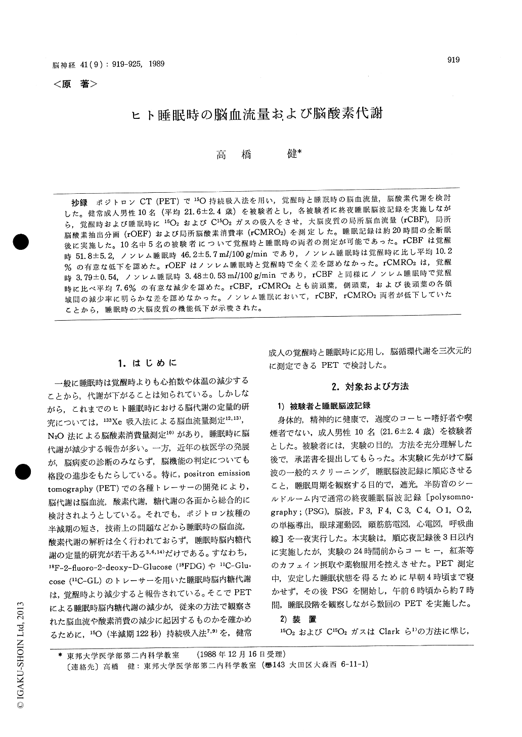
抄録 ポジトロンCT (PET)で15O持続吸入法を用い,覚醒時と睡眠時の脳血流量,脳酸素代謝を検討した。健常成人男性10名(平均21.6±2.4歳)を被験者とし,各被験者に終夜睡眠脳波記録を実施しながら,覚醒時および睡眠時に15O2およびC15O2ガスの吸入をさせ,大脳皮質の局所脳血流量(rCBF),局所脳酸素抽出分画(rOEF)および局所脳酸素消費率(rCMRO2)を測定した。睡眠記録は約20時間の全断眠後に実施した。10名中5名の被験者について覚醒時と睡眠時の両者の測定が可能であった。rCBFは覚醒時51.8±5.2,ノンレム睡眠時46.2±5.7ml/100g/minであり,ノンレム睡眠時は覚醒時に比し平均10.2%の有意な低下を認めた。rOEFはノンレム睡眠時と覚醒時で全く差を認めなかった。rCMRO2は,覚醒時3.79±0.54,ノンレム睡眠時3.48±O.53ml/100g/minであり,rCBFと同様にノンレム睡眠時で覚醒時に比べ平均7.6%の有意な減少を認めた。rCBF,rCMRO2とも前頭葉,側頭葉,および後頭葉の各領域間の減少率に明らかな差を認めなかった。ノンレム睡眠において,rCBF,rCMRO2両者が低下していたことから,睡眠時の大脳皮質の機能低下が示唆された。
Regional cerebral blood flow (rCBF), regional oxy-gen extraction fraction (rOEF) and regional cerebral metabolic rate for oxygen (rCMRO2) were measured using the continuous inhalation technique for 15O with positron emission tomography (PET) during both wakefulness and sleep. Ten paid volunteers, with a mean age of 21.6 yrs., were deprived of sleep for a period of approximately 20 hours, and the ex-periments were performed mostly in the morning. 15Oactivity of both whole blood and the plasma, pixel count of PET, total arterial blood oxygen content were used for analysis of rCBF, rOEF and rCMRO2. PET scannings were carried out mostly during the very light non-rapid eye movement (NREM) sleep, i. e. stage 1 and/or 2, and wakefulness. About 10 min-utes after the start of continuous inhalation of 15O gas, the 15O activity of the brain was found to be in a steady-state condition. During this steady-state condition, PET scannings were performed for about 10 minutes. Regions of interest, square in shape and having an area of 2.8 cm3, were set in each cortex on PET images of a horizontal cross-section of the brain, set at 45 mm above the orbitomeatal line. The rCBF and rCMROz were analysed in 5 of 10 male subjects during both wakefulness and NREM sleep, and only 3 were done during three sleep stages, in-cluding REM sleep. Levels of rCBF and rCMRO2 were found to be decreased in NREM sleep, and the decreasing rates were calculated at 10.2% and 7.6% from the level of wakefulness, respectively. There was no significant difference in the mean value of rOEF between wakefulness and NREM sleep. There were no significant regional differences found in the rate of decrease among the frontal, temporal and oc-cipital cortices. It was considered that the decrease of rCBF and rCMRO2 during NREM sleep suggested a decrease of the activity levels in the cerebral func-tions.

Copyright © 1989, Igaku-Shoin Ltd. All rights reserved.


