Japanese
English
- 有料閲覧
- Abstract 文献概要
- 1ページ目 Look Inside
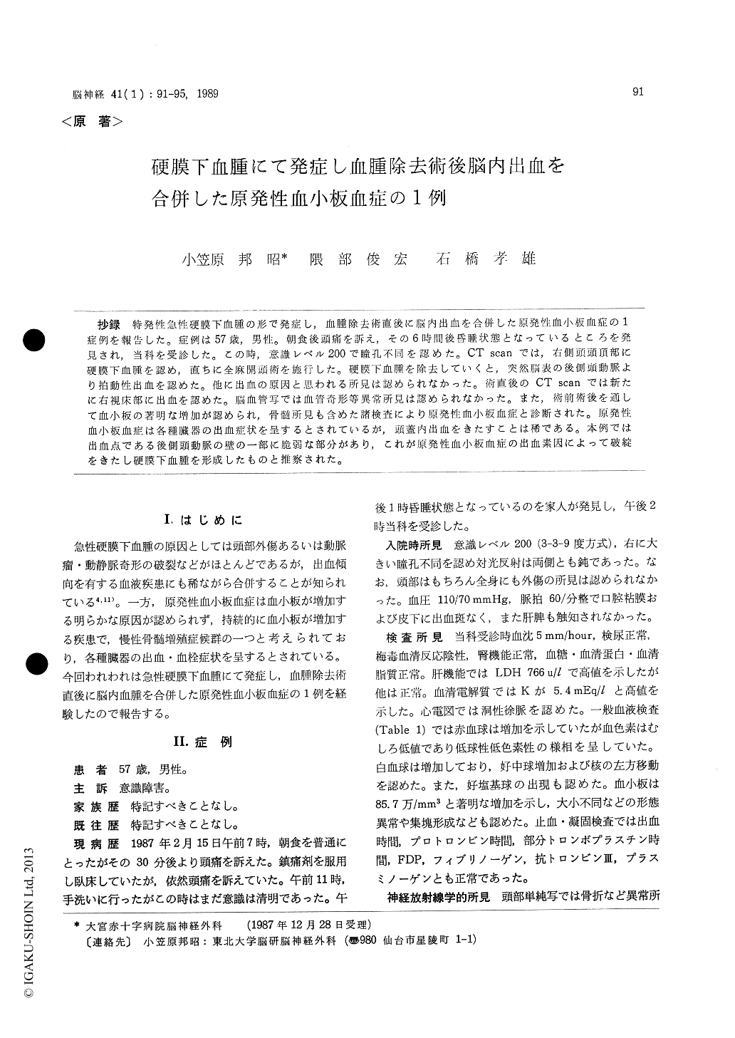
抄録 特発性急性硬膜下血腫の形で発症し,血腫除去術直後に脳内出血を合併した原発性血小板血症の1症例を報告した。症例は57歳,男性。朝食後頭痛を訴え,その6時間後昏睡状態となっているところを発見され,当科を受診した。この時,意識レベル200で瞳孔不同を認めた。CT scanでは,右側頭頭頂部に硬膜下血腫を認め,直ちに全麻開頭術を施行した。硬膜下血腫を除去していくと,突然脳表の後側頭動脈より拍動性出血を認めた。他に出血の原因と思われる所見は認められなかった。術直後のCT scanでは新たに右視床部に出血を認めた。脳血管写では血管奇形等異常所見は認められなかった。また,術前術後を通して血小板の著明な増加が認められ,骨髄所見も含めた諸検査により原発性血小板血症と診断された。原発性血小板血症は各種臓器の出血症状を呈するとされているが,頭蓋内出血をきたすことは稀である。本例では出血点である後側頭動脈の壁の一部に脆弱な部分があり,これが原発性血小板血症の出血素因によって破綻をきたし硬膜下血腫を形成したものと推察された。
A case of essential thrombocythemia (ET) as-ciated with subdural hematoma and postoperative intracerebral hemorrhage was reported. A 57-year-old man had complained headache in the morning. Six hours later he was found uncon-sciousness and soon he was brought to our hospi-tal. On admission he was comatose. There was no evidences of head injury and the X-rays were normal. A computed tomography (CT) scan re-vealed an acute subdural hematoma over the left temporoparietal region. Laboratory data revealed thrombocytosis of 85. 7x 10⁴/mm3 with increased red and white blood cell counts. Emergent right craniotomy was performed and a subdural clot was evacuated. Neither cortical damage nor vas-cular malformations were seen on the cortical surface. But a spurting cortical artery with a pin-hole could be seen. A postoptrative CT scan revealed an intracerebral hemorrhage deep in the right hemisphere. Cerebral angiograms revealed no vascular anomalies. Postoperatively, the platelet count remained high and laboratory data including bone marrow finding, neutrophil alkali-phosphatase score and chromosome analysis were consistent with the diagnosis of essential thrombocythemia. The mechanisms of subdural hematoma formation and postoperative intracerebral hemorrhage as-sociated with essential thrombocythemia were discussed.

Copyright © 1989, Igaku-Shoin Ltd. All rights reserved.


