Japanese
English
- 有料閲覧
- Abstract 文献概要
- 1ページ目 Look Inside
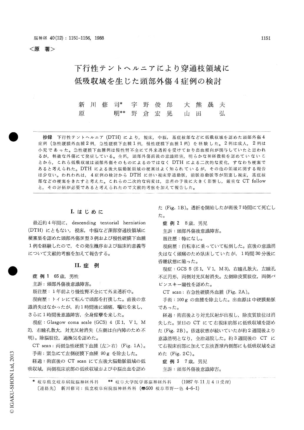
抄録 下行性テントヘルニア(DTH)により,視床,中脳,基底核部などに低吸収域を認めた頭部外傷4症例(急性硬膜外血腫2例,急性硬膜下血腫1例,慢性硬膜下血腫1例)を経験した。2例は成人,2例は小児であった。急性硬膜下血腫例は慢性腎不全にて外来透析を受けており出血傾向が関与していたと思われるが,軽徴な外傷にて発症している。全例,頭部外傷直後の意識障害,明らかな神経徴候を認めていないことから,これら低吸収域は頭部外傷そのものによるのではなくDTHによる二次的な変化,すなわち梗塞であると考えられた。DTHによる後大脳動脈領域の梗塞はよく知られているが,その他の領域に関する報告は少ない。われわれは,4症例の検討からDTHに伴い視床穿通動脈,前脈絡動脈等が閉塞し視床,甚底核部などの梗塞をきたすと考えた。これらの二次的な病変は,患者の予後に大きく影響し,厳重なCT followと,その評価が必要であると考えられたので文献的考察を加えて報告した。
Four cases with descending tentorial herniation(DTH) after head injury which showed thalamic,mesencephalic and basal ganglionic low density areas (LDAs) manifesting a infarction in postope-rative CT films are reported, and a possible mecha-nism are discussed in this paper. Case 1: Bilateral acute subdural hematoma with left DTH showed LDAs in the anterior part of the bilateral thalami, left occipital lobe and midbrain. The estimated occluded arteries included the anterior thlamoper-forating artery(AThA),posterior cerebral artery and midbrain perforator. Case 2: Right acute epidural hematoma with DTH showed LDAs in the anterior part of the right thalamus and in the left globus pallidus. The estimated occluded arteries included the AThA and anterior choroidal artery. Case 3: Right acute epidural hematoma with DTH showed LDAs in the anterior part of the left thalamus, and in the left midbrain tegmentum. The estimated occluded artery was the interpeduncular thalamo-perforating artery (IThA). Case 4: Right chronic subdural hematoma with DTH showed LDA main-ly in the left thalamus except for the superior thalamic region. The estimated occluded arteries included the AThA and/or IThA and thalamo-geniculate artery. Cases 1 and 4 were adult males and cases 2 and 3 were infant males, and the prog-nosis was good in the infant males, and poor in the adult males.
Each of the 4 cases showed no loss of conscious-ness just after the head injury while 3 out of them deteriorated within several hours, and one was a case of chronic subdural hematoma. Therefore, it was suggested that the thalamic, mesencephalic and basal ganglionic LDAs were not due to head inju-ry itself, but rather due to stretching, compression. and obstruction of the arteries due to a shift of the brainstem by DTH. These secondary lesions exert a grave influence on the prognosis, and a strict CT follow must be performed.

Copyright © 1988, Igaku-Shoin Ltd. All rights reserved.


