Japanese
English
- 有料閲覧
- Abstract 文献概要
- 1ページ目 Look Inside
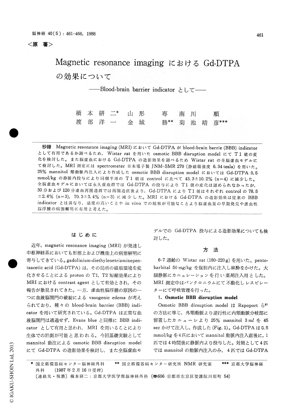
抄録 Magnetic resonance imaging (MRI)においてGd-DTPAがblood-brain barrie (BBB) indicatorとして有用であるか調べるため,Wistar ratを用いたosmotic BBB disruption modelにてT1値の変化を検討した。また脳虚血におけるGd-DTPAの造影効果を調べるためWistar ratの全脳虚血モデルにて検討した。MRI測定にはspectrometer日本電子製JNM-SMR 270(静磁場強度6.34tesla)を用いた。25%mannitol頸動脈内注入により作成したosmotic BBB disruption modelにおいてはGd-DTPA 0.5mmol/kgの静脈内投与により同側半球のT1値はcontrolに比べて43.3±10.2%(n=4)に減少した。全脳虚血モデルにおいては永久虚血群ではGd-DTPAの投与によりT1値の変化は認められなかったが,30分および120分虚血再開通群では再開通直後より,Gd-DTPAによりT1値はそれぞれcontrolの78.5±2.6%(n=3),70.3±3.4%(n=3)に減少した。MRIにおけるGd-DTPAの造影効果は従来のBBBindicatorとは異なり,感度の高いことやin vivoでの観察が可能なことより脳虚血巣の早期発見や虚血性脳浮腫の病態解明に有用と考えた。
Magnetic resonance image (MRI) has been applied on central nervous system to evaluate the anatomical and functional aspects. On the other hand, gadolinium-diethylenetriamine-pentaaceticacid (Gd-DTPA) is considered to be a valuable contrast agent on MRI. This paramagnetic com-pound shortens the magnetic relaxation times of surrounding hydrogen nuclei by altering local magnetic environments. A molecular weight of this compound was 590, therefore this does not pass through the normal blood-brain barrier (BBB), and it may possible to detect a breakdown of BBB in vivo.
In order to investigate the effect of Gd-DTPA as an indicator of BBB disruption on MRI, we measured T1 value of hemisphere of rat in osmotic BBB disruption model and experimental global cerebral ischemic model with and without Gd-DTPA. The MRI system employed was JNM-SMR 270 (JEOL) and the superconducting magnet was operated at a field strength of 6. 34 tesla. Gd-DTPA was injected intra-venously through femoral vein and its dose was 0.5 mmol/kg. Osmotic BBB dis-ruption model was made by intra-arterial injection of 25% mannitol through an internal carotid artery. Calculated T1 value of the ipsilateral hemisphere decreased with Gd-DTPA to 43.3±10.2% (n=4) of control value immediately after BBB disruption with mannitol administration but no change of T1 value was recognized with Gd-DTPA in rats which Gd-DTPA was injected 4 hours after BBB disrup-tion. Global ischemic insult was made by 3 vessel occlusion permanently and temporarily (30 or 120 minutes). In permanent occlusion models, the chan-ges of T1 value were not recognized with or without Gd-DTPA within 4-6 hours after occlusion. But in recirculation models after 30 minutes ischemia, the T1 value decreased to 78.5±2.6% (n=3) with Gd-DTPA immediately after recircula-tion, and the reduction of Ti value with Gd-DTPA was gradually decreased with the lapse of time after recirculation. In recirculation models after 120 minutes ischemia, the T1 value decreased 70. 3 ±3.4% (n=3) with Gd-DTPA immediately after recirculation, but the reduction of T1 value was not detected in rats were injected with Gd-DTPA 4 hours after recirculation.
In conclusion, the enhancement of T1 value with Gd-DTPA was recognized in osmotic BBB disrup-tion model and ischemic model. And this enhance-ment was observed immediately after recirculation in temporarily ischemic models. This suggests pos-sibility of early diagnosis of cerebral infarction with use of Gd-DTPA. Gd-DTPA on MRI was considered to be very sentitive and three dimen-sional BBB disrupted indicator in vivo, and to be feasible in evaluating the BBB function in ischemic edema.

Copyright © 1988, Igaku-Shoin Ltd. All rights reserved.


