Japanese
English
- 有料閲覧
- Abstract 文献概要
- 1ページ目 Look Inside
抄録 典型的な連合型視覚失認の1例を経験した。症例は61歳,男。頭部外傷を契機として,物体失認,失読,失書,相貌失認,色名呼称障害,外傷以後における言語的記憶障害と三次元空間における構成障害を認めた。X線CTおよびMRIから両側側頭後頭葉領域の白質(両側下縦束を含む)を中心とした病巣を確認した。物体失認の発現機序として,解剖学的には両側下縦束損傷に伴う半球内離断が,心理学的機序として視覚認知に関する立体的な視覚構成能力,さらにその三次元的な推察能力の脱落が視覚失認の発現に一因をなしている可能性が示唆された。
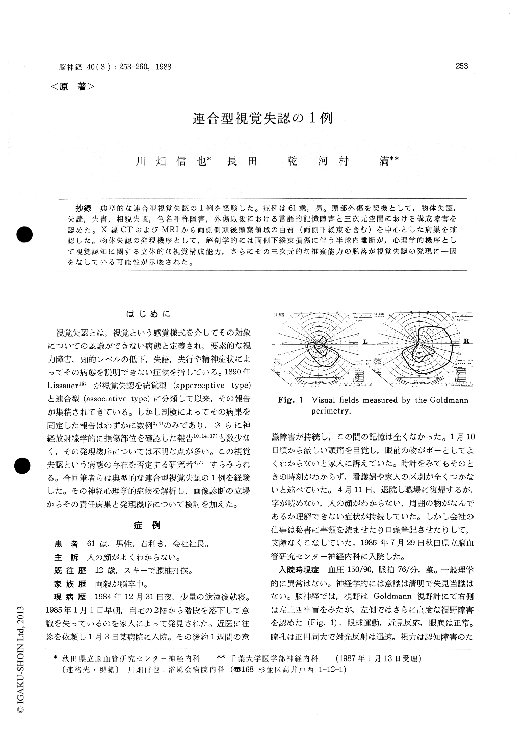
A 61 year-old right-handed man, a president of a company fell downstairs and was found unconscious on January 1, 1985. He was admitted to a hospital and he had been unresponsive for 10 days. As he recovered from the consciousness disturbance,he complained of difficulties in discrimi-nation of common objects and in recognition of familiar faces. The patient was admitted to our hospital for the closer examination on July 29, 1985. There was no abnormality in the general physical examination on admission. Neurological examinations revealed no significant deficits except visual field disturbance. In the results of WAIS, the verbal IQ was 108, but the performance IQ was unmeasurable because of a presence of object agnosia. His spontaneous speech was fluent without paraphasia, perseveration or dysprosody. His au-ditory comprehension of spoken language and repetition were spared. Neuropsychological ex-aminations revealed an impairment in the recent verbal memory, alexia, agraphia, object agnosia, color naming difficulty, prosopagnosia and visuo-spatial constructional disability. The patient ex-hibited three major neuropsychological chara-cteristics. Firstly, the recognition of the objects which were presented by the photograph or line drawing was more severely impaired than the real objects. Secondly, the object naming difficulty was more pronounced in the presentation at an unusual angle than in the free presentation. Thirdly, the patient made poor performance in reproduction of the models by photographic presentation as compared with the presentation of the real models in the three-dimensional block construction test. The CT scan performed on January 4, 1985 demonstrated subcortical hema-tomas with surrounding edema in the temporo-occipital regions of both hemispheres. No abnormal density was detected in either parietal lobe orcorpus callosum on the follow-up CT. Magnetic resonance imaging performed on May 17, 1986 demonstrated extensive low intensity lesions in the lingual, fusiform and posterior inferior tem-poral gyri of both hemispheres. The both inferior longitudinal fasciculi were also affected. In spite of the absence of elementary visual disturbance, aphasia or dementia, the patient showed a dis-ability in recognition or naming of the common objects. This type of disturbance in the visual recognition of objects could be differentiated from an apperceptic visual agnosia by the results that the copying and matching of objects were intact in this patient. The naming difficulty of the visually presented objects has also been described in patients with optic aphasia. When the patient with visual agnosia failed to name the objects, he could neither demonstrate their usage by pantomime nor point to them according to the verbal commands. In addition, he was unable to categorize the objects. These clinical evidences may support that this naming difficulty of the visually presented objects differs from that of the optic aphasia. His neuropsychological mani-festations seem best described as a typical form of the associative visual agnosia. The neuro-radiological findings suggest that the associative visual agnosia might be produced by an intra-hemispheric disconnection between the visual cor-tices and the temporal lobes, specifically the lim-bic system in which the visual memory is stored. In addition to this anatomical hypothesis, the visuo-constructional ability to convert the two-dimensional input to the three-dimensional const-ruction and the ability of three-dimensional ima-gination were impaired in this patient. These neuropsychological features were considered to participate in the genesis of the associative visual agnosia (psychological hypothesis).
Copyright © 1988, Igaku-Shoin Ltd. All rights reserved.