Japanese
English
- 有料閲覧
- Abstract 文献概要
- 1ページ目 Look Inside
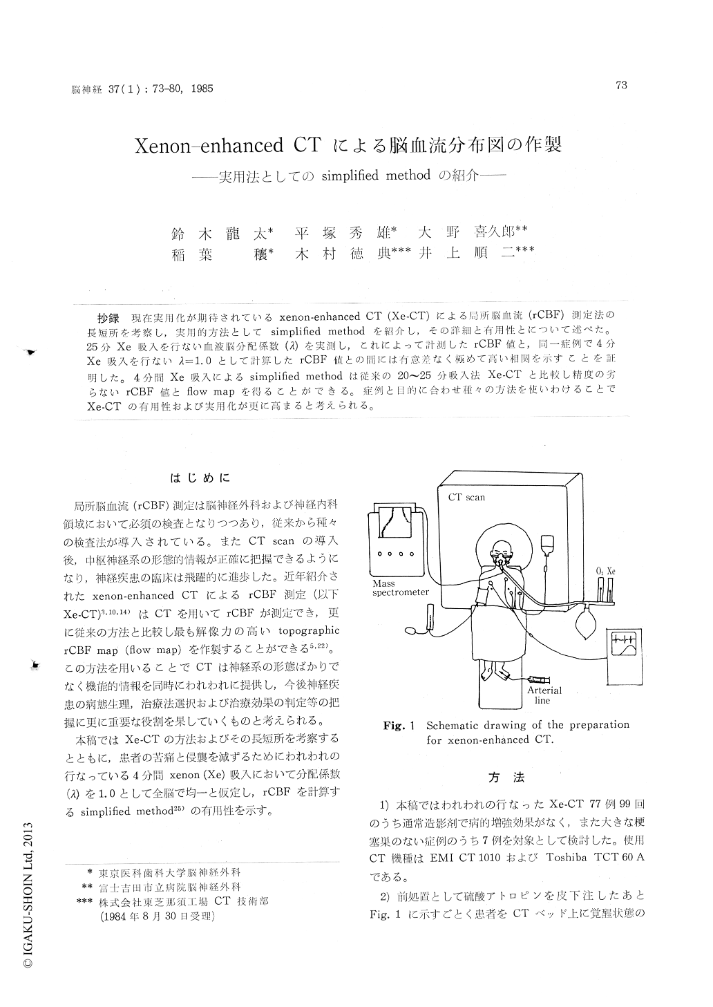
抄録 現在実用化が期待されているxenon-enhanced CT(Xe-CT)による局所脳血流(rCBF)測定法の長短所を考察し,実用的方法としてsimplified methodを紹介し,その詳細と有用性とについて述べた。25分Xe吸入を行ない血液脳分配係数(λ)を実測し,これによって計測したrCBF値と,同一症例で4分Xe吸入を行ないλ=1.0として計算したrCBF値との間には有意差なく極めて高い相関を示すことを証明した。4分間Xe吸入によるsimplified methodは従来の20〜25分吸入法Xe-CTと比較し精度の劣らないrCBF値とflow mapを得ることができる。症例と目的に合わせ種々の方法を使いわけることでXe-CTの有用性および実用化が更に高まると考えられる。
Measurement of reginal cerebral blood flow (rCBF) has become a routine examinations in the field of clinical neurology and neurosurgery. Therefore, various methods for measuring rCBF have been developed. Among them, xenon-enhanced CT method has many advantages compared with others. An obtainable topogaraphic flow map, high anatomical spatial resolution, and readily available instrument, namely CT scanner, are regarded as the advantages. On the one hand, anesthetic effects of xenon gas on the patients, taking a great expense for a single examination, and a significant dose of radiation delivered to the patients are thought to be the disadvantages. Due to the disadvantages, xenon-enhanced CT is regarded as an unpractical examination.
In the paper, we introduce a method with a brief xenon inhalation, named a simplified method, with which the disadvantages can be avoided. Conventional xenon-enhanced CT requires 20 to 25 minute inhalation of xenon gas in order to achieve a saturation of xenon in the cerebral tissue for a calculation of the partition coefficients (L). In the simplified method, instead of using the calculated L, L was given as 1.0 in all the cerebral regions and xenon gas was terminated at 4 minute inhalation. Four minute inhalation of xenon gas did not make any significant anesthetic effects on the patients nor the changes of physiological parameters, such as, PaCO2 and blood pressure. rCBF values calculated with the various durations of xenon inhalation using two different Ls : one was L calculated based on the saturated scan, and the other was L fixed as i. 0, revealed that theboth values were almost similar when 3 to 8 minute inhalation were chosen. Making a comparison between rCBF values obtained by the conventional method and those obtained by the simplified method, proved that they were statistically equal in the corresponding cerebral regions. The formulas used here are as follows:
1) integrated method,
(式省略)
2) autoradiographic method,
(式省略)
where Ci and Ca are the tissue and arterial bloodxenon concentrations respectively, K is the tissue build up flow rate, and f is the flow in the chosen compartments.
Generally we performed the simplified method and computed with the formula (1), and in some cases the conventional method was performed when the analyses of L was needed. The autoradiographic method is thought to be less reliable than the integrated method, however, with a single performance of xenon-enhanced CT, multiple flow maps are to be obtained by the autoradiographic method in order to avoid the unnecessary overirradiation.
Copyright © 1985, Igaku-Shoin Ltd. All rights reserved.